Pelvic cavity

Last updated
Pelvic cavity
Body Cavities Frontal view labeled.jpg
The various cavities of the human body as seen in a frontal projection, with the pelvic cavity labeled 4.
Body Cavities Lateral view labeled.jpg
The various cavities of the human body as seen in a lateral projection, with the pelvic cavity labeled 4.
Details
Artery Ovarian artery, Internal iliac artery, Median sacral artery
Vein Internal iliac vein, internal pudendal vein, vesical vein
Nerve Inferior hypogastric plexus
Lymph Primarily internal iliac lymph nodes
Identifiers
Latin cavitas pelvis
MeSH D057892
TA98 A02.5.02.002
TA2 1283
FMA 9738
Anatomical terminology
Gray241.png
Male pelvis.
Gray242.png
Female pelvis.

The pelvic cavity is a body cavity that is bounded by the bones of the pelvis. Its oblique roof is the pelvic inlet (the superior opening of the pelvis). Its lower boundary is the pelvic floor.

Contents

The pelvic cavity primarily contains the reproductive organs, urinary bladder, distal ureters, proximal urethra, terminal sigmoid colon, rectum, and anal canal. In females, the uterus, fallopian tubes, ovaries and upper vagina occupy the area between the other viscera. [1] [2]

The rectum is located at the back of the pelvis, in the curve of the sacrum and coccyx; the bladder is in front, behind the pubic symphysis. The pelvic cavity also contains major arteries, veins, muscles, and nerves. These structures coexist in a crowded space, and disorders of one pelvic component may impact upon another; for example, constipation may overload the rectum and compress the urinary bladder, or childbirth might damage the pudendal nerves and later lead to anal weakness.

Structure

Pelvis Pelvis.jpg
Pelvis

The pelvis has an anteroinferior, a posterior, and two lateral pelvic walls; and an inferior pelvic wall, also called the pelvic floor. [3] [4] The parietal peritoneum is attached here and to the abdominal wall. [5]

Lesser pelvis

The lesser pelvis (or "true pelvis") is the space enclosed by the pelvic girdle and below the pelvic brim: between the pelvic inlet and the pelvic floor. This cavity is a short, curved canal, deeper on its posterior than on its anterior wall. [1] Some sources consider this region to be the entirety of the pelvic cavity. Other sources define the pelvic cavity as the larger space including the greater pelvis, just above the pelvic inlet.

The lesser pelvis is bounded in front and below by the superior rami of the symphysis pubis; above and behind, by the sacrum and coccyx; and laterally, by a broad, smooth, quadrangular area of bone, corresponding to the inner surfaces of the body and superior ramus of the ischium, and the part of the ilium below the arcuate line. [1]

roof: pelvic brim [6]   
  posterior: sacrum, coccyx    lateral: obturator internus    anterior: pubic symphysis  
floor: pelvic floor     

The lesser pelvis contains the pelvic colon, rectum, bladder, and some of the sex organs. The rectum is at the back, in the curve of the sacrum and coccyx; the bladder is in front, behind the pubic symphysis. In females, the uterus and vagina occupy the interval between these viscera. [1]

The pelvic splanchnic nerves arising at S2–S4 are in the lesser pelvis.

Greater pelvis

The greater pelvis (or false pelvis) is the space enclosed by the pelvic girdle above and in front of the pelvic brim. It is bounded on either side by the ilium. In the front, it is incomplete, presenting a wide interval between the anterior borders of the ilia, which is filled by the muscles and fascia of the anterior abdominal wall; behind is a deep notch on either side between the ilium and the base of the sacrum that is filled by the thoracolumbar fascia and associated muscles. [1]

It is generally considered part of the abdominal cavity (which is why it is sometimes called the false pelvis). [7] Some sources consider this region part of the pelvic cavity,[ citation needed ] while others reframe the classification by calling the combination the abdominopelvic cavity.

The greater pelvis supports the intestines (specifically, the ileum and sigmoid colon), and transmits part of their weight to the anterior wall of the abdomen. [1]

The femoral nerve from L2–L4 is in the greater pelvis, but not in the lesser pelvis.

Ligaments

LigamentFromTo
broad ligament of the uterus uterusside of pelvis
* mesovarium ovary
* mesosalpinx Fallopian tube broad ligament of the uterus
* mesometrium
cardinal ligament cervix and vaginapelvic wall
ovarian ligament ovaryuterus
round ligament of the uterus ovarytravels through inguinal canal, ends at mons pubis
suspensory ligament of the ovary ovarypelvic wall

Arteries

Nerves

Measurements

The pelvis can be classified into four main types by measuring the pelvic diameters and conjugates at the pelvic inlet and outlet and as oblique diameters.

Female pelvic cavity Anatomical dissection of the pelvic cavity.jpg
Female pelvic cavity
Pelvic measurements [8]
MeasurementFromToLength
Transverse diameter
(of inlet)
Between extreme lateral points of pelvic inlet13.5–14 cm
Oblique diameter IRight sacroiliac joint Left iliopubic eminence12-12.5 cm
Oblique diameter IILeft sacroiliac jointRight iliopubic eminence11.5–12 cm
Anatomical conjugate
(true conjugate)
Pubic symphysisPromontory~12 cm
Obstetric conjugateRetropubic eminence
(posterior surface
of symphysis)
Promontory>10 cm
Diagonal conjugate*Inferior pubic ligamentPromontory11.5–12 cm
Straight conjugateLower border of symphysisTip of coccyx9.5–10 cm
Median conjugateLower border of symphysisLower border of sacrum11.5 cm
Transverse diameter
(of outlet)
Between ischial tuberosities 10–11 cm
Interspinous distanceBetween anterior superior iliac spines26 cm
(female)
Intercristal distanceBetween furthest lateral points of iliac crest 29 cm
(female)
External conjugateSpinous process of fifth lumbar vertebraUpper edge of symphysis~20 cm
Intertrochanteric distanceBetween femurs31 cm
Because the true conjugate cannot be measured directly, it is derived from the diagonal conjugate, which is measured through the vagina.

Additional images

Related Research Articles

<span class="mw-page-title-main">Levator ani</span> Broad, thin muscle group, situated on either side of the pelvis

The levator ani is a broad, thin muscle group, situated on either side of the pelvis. It is formed from three muscle components: the pubococcygeus, the iliococcygeus, and the puborectalis.

<span class="mw-page-title-main">Sacrum</span> Triangular-shaped bone at the bottom of the spine

The sacrum, in human anatomy, is a large, triangular bone at the base of the spine that forms by the fusing of the sacral vertebrae (S1–S5) between ages 18 and 30.

<span class="mw-page-title-main">Spinal nerve</span> Nerve that carries signals between the spinal cord and the body

A spinal nerve is a mixed nerve, which carries motor, sensory, and autonomic signals between the spinal cord and the body. In the human body there are 31 pairs of spinal nerves, one on each side of the vertebral column. These are grouped into the corresponding cervical, thoracic, lumbar, sacral and coccygeal regions of the spine. There are eight pairs of cervical nerves, twelve pairs of thoracic nerves, five pairs of lumbar nerves, five pairs of sacral nerves, and one pair of coccygeal nerves. The spinal nerves are part of the peripheral nervous system.

<span class="mw-page-title-main">Sigmoid colon</span> Section of the large intestine closest to the rectum and anus

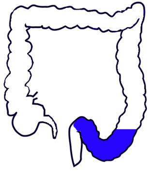
The sigmoid colon is the part of the large intestine that is closest to the rectum and anus. It forms a loop that averages about 35–40 centimetres (14–16 in) in length. The loop is typically shaped like a Greek letter sigma (ς) or Latin letter S. This part of the colon normally lies within the pelvis, but due to its freedom of movement it is liable to be displaced into the abdominal cavity.

<span class="mw-page-title-main">Internal pudendal artery</span> Blood vessel supplying blood to the external genitalia

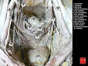
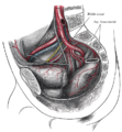
The internal pudendal artery is one of the three pudendal arteries. It branches off the internal iliac artery, and provides blood to the external genitalia.

<span class="mw-page-title-main">Sacral plexus</span> Nerve plexus

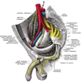
In human anatomy, the sacral plexus is a nerve plexus which provides motor and sensory nerves for the posterior thigh, most of the lower leg and foot, and part of the pelvis. It is part of the lumbosacral plexus and emerges from the lumbar vertebrae and sacral vertebrae (L4-S4). A sacral plexopathy is a disorder affecting the nerves of the sacral plexus, usually caused by trauma, nerve compression, vascular disease, or infection. Symptoms may include pain, loss of motor control, and sensory deficits.

<span class="mw-page-title-main">Internal iliac artery</span> Main artery of the pelvis

The internal iliac artery is the main artery of the pelvis.

<span class="mw-page-title-main">Sacrotuberous ligament</span>

The sacrotuberous ligament is situated at the lower and back part of the pelvis. It is flat, and triangular in form; narrower in the middle than at the ends.

<span class="mw-page-title-main">Superior hypogastric plexus</span>

The superior hypogastric plexus is a plexus of nerves situated on the vertebral bodies anterior to the bifurcation of the abdominal aorta. It bifurcates to form the left and the right hypogastric nerve. The SHP is the continuation of the abdominal aortic plexus.

<span class="mw-page-title-main">Pelvic brim</span>

The pelvic brim is the edge of the pelvic inlet. It is an approximately Mickey Mouse head-shaped line passing through the prominence of the sacrum, the arcuate and pectineal lines, and the upper margin of the pubic symphysis.

<span class="mw-page-title-main">Internal iliac vein</span> Large blood vessel of the pelvis

The internal iliac vein begins near the upper part of the greater sciatic foramen, passes upward behind and slightly medial to the internal iliac artery and, at the brim of the pelvis, joins with the external iliac vein to form the common iliac vein.

<span class="mw-page-title-main">Nerve to obturator internus</span> Human nerve

The nerve to obturator internus is a mixed nerve providing motor innervation to the obturator internus muscle and gemellus superior muscle, and sensory innervation to the hip joint. It is a branch of the sacral plexus. It is one of the group of deep gluteal nerves.

<span class="mw-page-title-main">Inferior hypogastric plexus</span>

The inferior hypogastric plexus is a network of nerves that supplies the organs of the pelvic cavity. The inferior hypogastric plexus gives rise to the prostatic plexus in males and the uterovaginal plexus in females.

Pelvic splanchnic nerves or nervi erigentes are splanchnic nerves that arise from sacral spinal nerves S2, S3, S4 to provide parasympathetic innervation to the organs of the pelvic cavity.

<span class="mw-page-title-main">Urogenital triangle</span> Anterior part of the perineum

The urogenital triangle is the anterior part of the perineum. In female mammals, it contains the vagina and associated parts of the internal genitalia.

<span class="mw-page-title-main">Anal triangle</span> Posterior part of the perineum

The anal triangle is the posterior part of the perineum. It contains the anal canal.

<span class="mw-page-title-main">Hypogastric nerve</span> Human nerves

The hypogastric nerves are the continuation of the superior hypogastric plexus that descend into the pelvis anterior the sacrum and become the inferior hypogastric plexuses on either side of pelvic organs. The hypogastric nerves serve as a pathway for autonomic fibers to communicate between the lower abdomen and pelvis.

<span class="mw-page-title-main">Outline of human anatomy</span> Overview of and topical guide to human anatomy

The following outline is provided as an overview of and topical guide to human anatomy:

<span class="mw-page-title-main">Hip bone</span> Bone of the pelvis

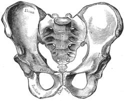
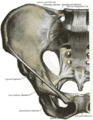
The hip bone is a large flat bone, constricted in the center and expanded above and below. In some vertebrates it is composed of three parts: the ilium, ischium, and the pubis.

<span class="mw-page-title-main">Pelvis</span> Lower torso of the human body

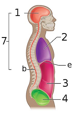
The pelvis is the lower part of the trunk, between the abdomen and the thighs, together with its embedded skeleton.

References

  1. 1 2 3 4 5 6 PD-icon.svgThis article incorporates text in the public domain from the 20th edition of Gray's Anatomy (1918)
  2. Drake R, Vogl AW, Mitchell AW (2018). "5 Pelvis and Perineum". Gray's Basic Anatomy E-Book. Elsevier. pp. 220–227. ISBN   978-0-323-50850-6.{{cite book}}: CS1 maint: multiple names: authors list (link)
  3. Moore, Keith L. et al. (2010) Clinically Oriented Anatomy 6th Ed, ch. 3 Pelvis and perineum, p. 339
  4. Richard S. Snell Clinical Anatomy By Regions, Pelvic cavity p. 242
  5. Tank, P. (2013) Grants Dissector 15th ed., ch. 4 The abdomen, p. 99
  6. "Anatomy of the Female Pelvis – D. El-Mowafi" . Retrieved 2007-12-03.
  7. Drake et al. (2009) Grays Anatomy for Students, 2nd Edition, ch. 5 Pelvis and perineumgeneral description, p. 406
  8. Platzer, Werner (2004). Color Atlas of Human Anatomy, Vol. 1: Locomotor System (5th ed.). Thieme. p. 190. ISBN   3-13-533305-1.