Body cavity

Last updated
Body cavity
Scheme body cavities-en.svg
Cross-section showing cavities in the human body, the dorsal and ventral body cavities labelled.
Annelid redone w white background.svg
Cross-section of an oligochaete worm. The worm's body cavity surrounds the central typhlosole.
Identifiers
FMA 85006
Anatomical terminology

A body cavity is any space or compartment, or potential space, in an animal body. Cavities accommodate organs and other structures; cavities as potential spaces contain fluid.

Contents

The two largest human body cavities are the ventral body cavity, and the dorsal body cavity. In the dorsal body cavity the brain and spinal cord are located.

The membranes that surround the central nervous system organs (the brain and the spinal cord, in the cranial and spinal cavities) are the three meninges. The differently lined spaces contain different types of fluid. In the meninges for example the fluid is cerebrospinal fluid; in the abdominal cavity the fluid contained in the peritoneum is a serous fluid.

In amniotes and some invertebrates the peritoneum lines their largest body cavity called the coelom.

Mammals

Mammalian embryos develop two body cavities: the intraembryonic coelom and the extraembryonic coelom (or chorionic cavity). The intraembryonic coelom is lined by somatic and splanchnic lateral plate mesoderm, while the extraembryonic coelom is lined by extraembryonic mesoderm. The intraembryonic coelom is the only cavity that persists in the mammal at term, which is why its name is often contracted to simply coelomic cavity. Subdividing the coelomic cavity into compartments, for example, the pericardial cavity / pericardium, where the heart develops, simplifies discussion of the anatomies of complex animals.

Cavitation in the early embryo is the process of forming the blastocoel, the fluid-filled cavity defining the blastula stage in non-mammals, or the blastocyst in mammals.

Human body cavities

The dorsal (posterior) cavity and the ventral (anterior) cavity are the largest body compartments.

The dorsal body cavity includes the cranial cavity, enclosed by the skull and contains the brain, and the spinal cavity, enclosed by the spine, and contains the spinal cord. [1]

The ventral body cavity includes the thoracic cavity, enclosed by the ribcage and contains the lungs and heart; and the abdominopelvic cavity. The abdominopelvic cavity can be divided into the abdominal cavity, enclosed by the ribcage and pelvis and contains the kidneys, ureters, stomach, intestines, liver, gallbladder, and pancreas; and the pelvic cavity, enclosed by the pelvis and contains bladder, anus and reproductive system. [1]

Human body cavities and membranes
Name of cavityPrincipal contentsMembranous lining
Dorsal body cavity Cranial cavity Brain Meninges
Vertebral canal Spinal cord Meninges
Ventral body cavity Thoracic cavity Heart, Lungs Pericardium
Pleural cavity
Abdominopelvic cavity Abdominal cavity Digestive organs, spleen, kidneys Peritoneum
Pelvic cavity Bladder, reproductive organs Peritoneum

Ventral body cavity

The ventral cavity has two main subdivisions: the thoracic cavity and the abdominopelvic cavity. The thoracic cavity is the more superior subdivision of the ventral cavity, and is enclosed by the rib cage. The thoracic cavity contains the lungs surrounded by the pleural cavity, and the heart surrounded by the pericardial cavity, located in the mediastinum. The diaphragm forms the floor of the thoracic cavity and separates it from the more inferior abdominopelvic cavity.

The abdominopelvic cavity is the largest cavity in the body occupying the entire lower half of the trunk. Although no membrane physically divides the abdominopelvic cavity, it can be useful to distinguish between the abdominal cavity, and the pelvic cavity. The abdominal cavity occupies the entire lower half of the trunk, anterior to the spine, and houses the organs of digestion. Just under the abdominal cavity, anterior to the buttocks, is the pelvic cavity. The pelvic cavity is funnel shaped, and is located inferior and anterior to the abdominal cavity, and houses the organs of reproduction. [2]

Dorsal body cavity

The dorsal body cavity contains the cranial cavity, and the spinal cavity. The cranial cavity is a large, bean-shaped cavity filling most of the upper skull where the brain is located. The spinal cavity is the very narrow, thread-like cavity running from the cranial cavity down the entire length of the spinal cord.

In the dorsal cavity, the cranial cavity houses the brain, and the spinal cavity encloses the spinal cord. Just as the brain and spinal cord make up a continuous, uninterrupted structure, the cranial and spinal cavities that house them are also continuous. The brain and spinal cord are protected by the bones of the skull and vertebral column and by cerebrospinal fluid, a colorless fluid produced by the brain, which cushions the brain and spinal cord within the dorsal body cavity. [2]

Development

At the end of the third week of gestation, the neural tube, which is a fold of one of the layers of the trilaminar germ disc, called the ectoderm, appears. This layer elevates and closes dorsally, while the gut tube rolls up and closes ventrally to create a "tube on top of a tube." The mesoderm, which is another layer of the trilaminar germ disc, holds the tubes together and the lateral plate mesoderm, the middle layer of the germ disc, splits to form a visceral layer associated with the gut and a parietal layer, which along with the overlying ectoderm, forms the lateral body wall. The space between the visceral and parietal layers of lateral plate mesoderm is the primitive body cavity. When the lateral body wall folds, it moves ventrally and fuses at the midline. The body cavity closes, except in the region of the connecting stalk. Here, the gut tube maintains an attachment to the yolk sac. The yolk sac is a membranous sac attached to the embryo, which provides nutrients and functions as the circulatory system of the very early embryo. [3]

The lateral body wall folds, pulling the amnion in with it so that the amnion surrounds the embryo and extends over the connecting stalk, which becomes the umbilical cord, which connects the fetus with the placenta. If the ventral body wall fails to close, ventral body wall defects can result, such as ectopia cordis, a congenital malformation in which the heart is abnormally located outside the thorax. Another defect is gastroschisis, a congenital defect in the anterior abdominal wall through which the abdominal contents freely protrude. Another possibility is bladder exstrophy, in which part of the urinary bladder is present outside the body. In normal circumstances, the parietal mesoderm will form the parietal layer of serous membranes lining the outside (walls) of the peritoneal, pleural, and pericardial cavities. The visceral layer will form the visceral layer of the serous membranes covering the lungs, heart, and abdominal organs. These layers are continuous at the root of each organ as the organs lie in their respective cavities. The peritoneum, a serum membrane that forms the lining of the abdominal cavity, forms in the gut layers and in places mesenteries extend from the gut as double layers of peritoneum. Mesenteries provide a pathway for vessels, nerves, and lymphatics to the organs. Initially, the gut tube from the caudal end of the foregut to the end of the hindgut is suspended from the dorsal body wall by dorsal mesentery. Ventral mesentery, derived from the septum transversum, exists only in the region of the terminal part of the esophagus, the stomach, and the upper portion of the duodenum. [4]

Function

These cavities contain and protect delicate internal organs, and the ventral cavity allows for significant changes in the size and shape of the organs as they perform their functions.

Anatomical structures are often described in terms of the cavity in which they reside. The body maintains its internal organization by means of membranes, sheaths, and other structures that separate compartments.

The lungs, heart, stomach, and intestines, for example, can expand and contract without distorting other tissues or disrupting the activity of nearby organs. [2] The ventral cavity includes the thoracic and abdominopelvic cavities and their subdivisions. The dorsal cavity includes the cranial and spinal cavities. [2]

Other animals

Organisms can be also classified according to the type of body cavity they possess, such as pseudocoelomates and protostome coelomates. [5]

Coelom

In amniotes and some invertebrates, the coelom is the large cavity lined by mesothelium, an epithelium derived from mesoderm. Organs formed inside the coelom can freely move, grow, and develop independently of the body wall while fluid in the peritoneum cushions and protects them from shocks.

Arthropods and most molluscs have a reduced (but still true) coelom, the hemocoel (of an open circulatory system) and the smaller gonocoel (a cavity that contains the gonads). Their hemocoel is often derived from the blastocoel.

See also

Related Research Articles

<span class="mw-page-title-main">Mesoderm</span> Middle germ layer of embryonic development

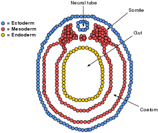
The mesoderm is the middle layer of the three germ layers that develops during gastrulation in the very early development of the embryo of most animals. The outer layer is the ectoderm, and the inner layer is the endoderm.

<span class="mw-page-title-main">Peritoneum</span> Serous membrane that forms lining of abdominal cavity or coelom

The peritoneum is the serous membrane forming the lining of the abdominal cavity or coelom in amniotes and some invertebrates, such as annelids. It covers most of the intra-abdominal organs, and is composed of a layer of mesothelium supported by a thin layer of connective tissue. This peritoneal lining of the cavity supports many of the abdominal organs and serves as a conduit for their blood vessels, lymphatic vessels, and nerves.

<span class="mw-page-title-main">Abdominal cavity</span> Body cavity in the abdominal area

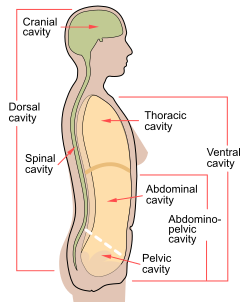
The abdominal cavity is a large body cavity in humans and many other animals that contain organs. It is a part of the abdominopelvic cavity. It is located below the thoracic cavity, and above the pelvic cavity. Its dome-shaped roof is the thoracic diaphragm, a thin sheet of muscle under the lungs, and its floor is the pelvic inlet, opening into the pelvis.

<span class="mw-page-title-main">Coelom</span> The main body cavity in many animals

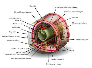
The coelom is the main body cavity in many animals and is positioned inside the body to surround and contain the digestive tract and other organs. In some animals, it is lined with mesothelium. In other animals, such as molluscs, it remains undifferentiated. In the past, and for practical purposes, coelom characteristics have been used to classify bilaterian animal phyla into informal groups.

<span class="mw-page-title-main">Pleural cavity</span> Thin fluid-filled space between the two pulmonary pleurae (visceral and parietal) of each lung

The pleural cavity, pleural space, or interpleural space is the potential space between the pleurae of the pleural sac that surrounds each lung. A small amount of serous pleural fluid is maintained in the pleural cavity to enable lubrication between the membranes, and also to create a pressure gradient.

<span class="mw-page-title-main">Mesothelium</span> Membrane lining body cavities

The mesothelium is a membrane composed of simple squamous epithelial cells of mesodermal origin, which forms the lining of several body cavities: the pleura, peritoneum and pericardium.

<span class="mw-page-title-main">Mesentery</span> Contiguous fold of tissues that supports the intestines

The mesentery is an organ that attaches the intestines to the posterior abdominal wall and is formed by the double fold of peritoneum. It helps in storing fat and allowing blood vessels, lymphatics, and nerves to supply the intestines, among other functions.

<span class="mw-page-title-main">Omphalocele</span> Rare abdominal wall defect in which internal organs remain outside of the abdomen in a sac

Omphalocele or omphalocoele also called exomphalos, is a rare abdominal wall defect. Beginning at the 6th week of development, rapid elongation of the gut and increased liver size reduces intra abdominal space, which pushes intestinal loops out of the abdominal cavity. Around 10th week, the intestine returns to the abdominal cavity and the process is completed by the 12th week. Persistence of intestine or the presence of other abdominal viscera in the umbilical cord results in an omphalocele.

<span class="mw-page-title-main">Yolk sac</span> Membranous sac attached to an embryo

The yolk sac is a membranous sac attached to an embryo, formed by cells of the hypoblast layer of the bilaminar embryonic disc. This is alternatively called the umbilical vesicle by the Terminologia Embryologica (TE), though yolk sac is far more widely used. In humans, the yolk sac is important in early embryonic blood supply, and much of it is incorporated into the primordial gut during the fourth week of embryonic development.

<span class="mw-page-title-main">Serous membrane</span> Smooth coating lining contents and inner walls of body cavities

The serous membrane is a smooth tissue membrane of mesothelium lining the contents and inner walls of body cavities, which secrete serous fluid to allow lubricated sliding movements between opposing surfaces. The serous membrane that covers internal organs is called visceral, while the one that covers the cavity wall is called parietal. For instance the parietal peritoneum is attached to the abdominal wall and the pelvic walls. The visceral peritoneum is wrapped around the visceral organs. For the heart, the layers of the serous membrane are called parietal and visceral pericardium. For the lungs they are called parietal and visceral pleura. The visceral serosa of the uterus is called the perimetrium. The potential space between two opposing serosal surfaces is mostly empty except for the small amount of serous fluid.

<span class="mw-page-title-main">Abdomen</span> Part of the body between the chest and pelvis

The abdomen is the part of the body between the thorax (chest) and pelvis, in humans and in other vertebrates. The abdomen is the front part of the abdominal segment of the torso. The area occupied by the abdomen is called the abdominal cavity. In arthropods, it is the posterior tagma of the body; it follows the thorax or cephalothorax.

<span class="mw-page-title-main">Cranial cavity</span> Space inside the skull formed by eight cranial bones known as the neurocranium

The cranial cavity, also known as intracranial space, is the space within the skull that accommodates the brain. The skull minus the mandible is called the cranium. The cavity is formed by eight cranial bones known as the neurocranium that in humans includes the skull cap and forms the protective case around the brain. The remainder of the skull is called the facial skeleton. Meninges are protective membranes that surround the brain to minimize damage to the brain in the case of head trauma. Meningitis is the inflammation of meninges caused by bacterial or viral infections.

<span class="mw-page-title-main">Septum transversum</span>

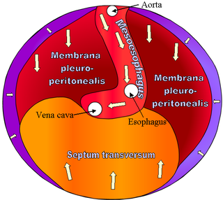
The septum transversum is a thick mass of cranial mesenchyme, formed in the embryo, that gives rise to parts of the thoracic diaphragm and the ventral mesentery of the foregut in the developed human being and other mammals.

<span class="mw-page-title-main">Lateral plate mesoderm</span>

The lateral plate mesoderm is the mesoderm that is found at the periphery of the embryo. It is to the side of the paraxial mesoderm, and further to the axial mesoderm. The lateral plate mesoderm is separated from the paraxial mesoderm by a narrow region of intermediate mesoderm. The mesoderm is the middle layer of the three germ layers, between the outer ectoderm and inner endoderm.

<span class="mw-page-title-main">Bilaminar embryonic disc</span>

The bilaminar embryonic disc, bilaminar blastoderm or embryonic disc is the distinct two-layered structure of cells formed in an embryo. In the development of the human embryo this takes place by day eight. It is formed when the inner cell mass, also known as the embryoblast, forms a bilaminar disc of two layers, an upper layer called the epiblast and a lower layer called the hypoblast, which will eventually form into fetus. These two layers of cells are stretched between two fluid-filled cavities at either end: the primitive yolk sac and the amniotic sac.

<span class="mw-page-title-main">Human embryonic development</span> Development and formation of the human embryo

Human embryonic development or human embryogenesis is the development and formation of the human embryo. It is characterised by the processes of cell division and cellular differentiation of the embryo that occurs during the early stages of development. In biological terms, the development of the human body entails growth from a one-celled zygote to an adult human being. Fertilization occurs when the sperm cell successfully enters and fuses with an egg cell (ovum). The genetic material of the sperm and egg then combine to form the single cell zygote and the germinal stage of development commences. Embryonic development in the human, covers the first eight weeks of development; at the beginning of the ninth week the embryo is termed a fetus. The eight weeks has 23 stages.

In the development of the human embryo, the intraembryonic coelom is a portion of the conceptus forming in the mesoderm during the third week of development. During the third week of development, the lateral plate mesoderm splits into a dorsal somatic mesoderm (somatopleure) and a ventral splanchnic mesoderm (splanchnopleure). The resulting cavity between the somatopleure and splanchnopleure is called the intraembryonic coelom. This space will give rise to the thoracic and abdominal cavities. The coelomic spaces in the lateral mesoderm and cardiogenic area are isolated. The isolated coelom begins to organize into a horseshoe shape. The spaces soon join together and form a single horseshoe-shaped cavity: the intraembryonic coelom. It then separates the mesoderm into two layers.

The development of the digestive system in the human embryo concerns the epithelium of the digestive system and the parenchyma of its derivatives, which originate from the endoderm. Connective tissue, muscular components, and peritoneal components originate in the mesoderm. Different regions of the gut tube such as the esophagus, stomach, duodenum, etc. are specified by a retinoic acid gradient that causes transcription factors unique to each region to be expressed. Differentiation of the gut and its derivatives depends upon reciprocal interactions between the gut endoderm and its surrounding mesoderm. Hox genes in the mesoderm are induced by a Hedgehog signaling pathway secreted by gut endoderm and regulate the craniocaudal organization of the gut and its derivatives. The gut system extends from the oropharyngeal membrane to the cloacal membrane and is divided into the foregut, midgut, and hindgut.

<span class="mw-page-title-main">Mesentery (zoology)</span> Membrane inside the body cavity of an animal

In zoology, a mesentery is a membrane inside the body cavity of an animal. The term identifies different structures in different phyla: in vertebrates it is a double fold of the peritoneum enclosing the intestines; in other organisms it forms complete or incomplete partitions of the body cavity, whether that is the coelom or, as in the Anthozoa, the gastrovascular cavity.

<span class="mw-page-title-main">Pulmonary pleurae</span> Serous membrane that lines the wall of the thoracic cavity and the surface of the lung

The pulmonary pleurae are the two opposing layers of serous membrane overlying the lungs, mediastinum and the inside surfaces of the surrounding chest walls.

References

This Wikipedia entry incorporates text from the freely licensed Connexions edition of Anatomy & Physiology text-book by OpenStax College

  1. 1 2 Ehrlich, A.; Schroeder, C.L. (2009), "The Human Body in Health and Disease", Introduction to Medical Terminology (Second ed.), Independence, KY: Delmar Cengage Learning, pp. 21–36
  2. 1 2 3 4 "Anatomy & Physiology". Openstax college at Connexions. Retrieved November 16, 2013.
  3. Sadler (2012). LANGMAN Embriología médica. Vol. I (12 ed.). Philadelphia, PA: The Point.
  4. Tortora, Gerard; Derrickson, Bryan (2008). Principios de anatomía y fisiología. Vol. I (11 ed.). Buenos Aires: Panamericana.
  5. "Animals III — Pseudocoelomates and Protostome Coelomates". Archived from the original on 2009-04-06.