Ectoderm

Last updated
Ectoderm
Ectoderm.png
Organs derived from ectoderm.
Gray11.png
Section through embryonic disk of Vespertilio murinus .
Details
Days16
Identifiers
MeSH D004475
FMA 69070
Anatomical terminology

The ectoderm is one of the three primary germ layers formed in early embryonic development. It is the outermost layer, and is superficial to the mesoderm (the middle layer) and endoderm (the innermost layer). [1] It emerges and originates from the outer layer of germ cells. The word ectoderm comes from the Greek ektos meaning "outside", and derma meaning "skin". [2]

Contents

Generally speaking, the ectoderm differentiates to form epithelial and neural tissues (spinal cord, nerves and brain). This includes the skin, linings of the mouth, anus, nostrils, sweat glands, hair and nails, [3] and tooth enamel. Other types of epithelium are derived from the endoderm. [3]

In vertebrate embryos, the ectoderm can be divided into two parts: the dorsal surface ectoderm also known as the external ectoderm, and the neural plate, which invaginates to form the neural tube and neural crest. [4] The surface ectoderm gives rise to most epithelial tissues, and the neural plate gives rise to most neural tissues. For this reason, the neural plate and neural crest are also referred to as the neuroectoderm.

History

Heinz Christian Pander, a Baltic German–Russian biologist, has been credited for the discovery of the three germ layers that form during embryogenesis. Pander received his doctorate in zoology from the University of Würzburg in 1817. He began his studies in embryology using chicken eggs, which allowed for his discovery of the ectoderm, mesoderm and endoderm. Due to his findings, Pander is sometimes referred to as the "founder of embryology".

Pander's work of the early embryo was continued by a PrussianEstonian biologist named Karl Ernst von Baer. Baer took Pander's concept of the germ layers and through extensive research of many different types of species, he was able to extend this principle to all vertebrates. Baer also received credit for the discovery of the blastula. Baer published his findings, including his germ layer theory, in a textbook which translates to On the Development of Animals which he released in 1828. [5]

Differentiation

Initial appearance

The ectoderm can first be observed in amphibians and fish during the later stages of gastrulation. At the start of this process, the developing embryo has divided into many cells, forming a hollow ball called the blastula. The blastula is polar, and its two halves are called the animal hemisphere and vegetal hemisphere. It is the animal hemisphere will eventually become the ectoderm. [2]

Early development

Like the other two germ layers – i.e., the mesoderm and endoderm – the ectoderm forms shortly after fertilization, after which rapid cell division begins. The position of the ectoderm relative to the other germ layers of the embryo is governed by "selective affinity", meaning that the inner surface of the ectoderm has a strong (positive) affinity for the mesoderm, and a weak (negative) affinity for the endoderm layer. [6] This selective affinity changes during different stages of development. The strength of the attraction between two surfaces of two germ layers is determined by the amount and type of cadherin molecules present on the cells' surface. For example, the expression of N-cadherin is crucial to maintaining separation of precursor neural cells from precursor epithelial cells. [2] Likewise, while the surface ectoderm becomes the epidermis, [6] the neuroectoderm is induced along the neural pathway by the notochord, which is typically positioned above it. [2] [4]

Gastrulation

During the process of gastrulation, bottle cells invaginate on the dorsal surface of the blastula to form the blastopore. The cells continue to extend inward and migrate along the inner wall of the blastula to form a fluid-filled cavity called the blastocoel. The once superficial cells of the animal pole are destined to become the cells of the middle germ layer called the mesoderm. Through the process of radial extension, cells of the animal pole that were once several layers thick divide to form a thin layer. At the same time, when this thin layer of dividing cells reaches the dorsal lip of the blastopore, another process occurs termed convergent extension. During convergent extension, cells that approach the lip intercalate mediolaterally, in such a way that cells are pulled over the lip and inside the embryo. These two processes allow for the prospective mesoderm cells to be placed between the ectoderm and the endoderm. Once convergent extension and radial intercalation are underway, the rest of the vegetal pole, which will become endoderm cells, is completely engulfed by the prospective ectoderm, as these top cells undergo epiboly, where the ectoderm cells divide in a way to form one layer. This creates a uniform embryo composed of the three germ layers in their respective positions. [2]

Later development

Once the three germ layers have been established, cellular differentiation can occur. The first major process here is neurulation, wherein the ectoderm differentiates to form the neural tube, neural crest cells and the epidermis. Each of these three components will give rise to a particular complement of cells. The neural tube cells give rise to the central nervous system, neural crest cells give rise to the peripheral and enteric nervous system, melanocytes, and facial cartilage, and the epidermal region will give rise to the epidermis, hair, nails, sebaceous glands, olfactory and oral epithelium, and eyes. [2]

Neurulation

Neurulation occurs in two parts, primary and secondary neurulation. Both processes position neural crest cells between a superficial epidermal layer and the deep neural tube. During primary neurulation, the notochord cells of the mesoderm signal the adjacent, superficial ectoderm cells to reposition themselves into a columnar pattern to form cells of the ectodermal neural plate. [7] As the cells continue to elongate, a group of cells immediately above the notochord change their shape, forming a wedge in the ectodermal region. These special cells are called medial hinge cells (MHPs). As the ectoderm continues to elongate, the ectodermal cells of the neural plate fold inward. The inward folding of the ectoderm by virtue of mainly cell division continues until another group of cells forms within the neural plate. These cells are termed dorsolateral hinge cells (DLHPs), and, once formed, the inward folding of the ectoderm stops. The DLHP cells function in a similar fashion as MHP cells regarding their wedge like shape, however, the DLHP cells result in the ectoderm converging. This convergence is led by ectodermal cells above the DLHP cells known as the neural crest. The neural crest cells eventually pull the adjacent ectodermal cells together, which leaves neural crest cells between the prospective epidermis and hollow, neural tube. [2]

Organogenesis

Ectodermal specification EctodermalSpecification.png
Ectodermal specification

All of the organs that rise from the ectoderm such as the nervous system, teeth, hair and many exocrine glands, originate from two adjacent tissue layers: the epithelium and the mesenchyme. [8] Several signals mediate the organogenesis of the ectoderm such as: FGF, TGFβ, Wnt, and regulators from the hedgehog family. The specific timing and manner that the ectodermal organs form is dependent on the invagination of the epithelial cells. [9] FGF-9 is an important factor during the initiation of tooth germ development. The rate of epithelial invagination in significantly increased by action of FGF-9, which is only expressed in the epithelium, and not in the mesenchyme. FGF-10 helps to stimulate epithelial cell proliferation, in order make larger tooth germs. Mammalian teeth develop from ectoderm derived from the mesenchyme: oral ectoderm and neural crest. The epithelial components of the stem cells for continuously growing teeth form from tissue layers called the stellate reticulum and the suprabasal layer of the surface ectoderm. [9]

Clinical significance

Ectodermal dysplasia

Ectodermal dysplasia is a rare but severe condition where the tissue groups (specifically teeth, skin, hair, nails and sweat glands) derived from the ectoderm undergo abnormal development. This is a diffuse term, as there are over 170 subtypes of ectodermal dysplasia. It has been accepted that the disease is caused by a mutation or a combination of mutations in certain genes. Research of the disease is ongoing, as only a fraction of the mutations involved with an ectodermal dysplasia subtype have been identified. [10]

Dental abnormalities in a 5-year-old girl from northern Sweden who suffered from various symptoms of autosomal dominant hypohidrotic ectodermal dysplasia (HED) a) Intraoral view. Note that the upper incisors have been restored with composite material to disguise their original conical shape. b) Orthopantomogram showing absence of ten primary and eleven permanent teeth in the jaws of the same individual. Dental abnormalities in a 5-year-old girl from north Sweden family who suffered from various symptoms of autosomal dominant hypohidrotic ectodermal dysplasia (HED).jpg
Dental abnormalities in a 5-year-old girl from northern Sweden who suffered from various symptoms of autosomal dominant hypohidrotic ectodermal dysplasia (HED) a) Intraoral view. Note that the upper incisors have been restored with composite material to disguise their original conical shape. b) Orthopantomogram showing absence of ten primary and eleven permanent teeth in the jaws of the same individual.

Hypohidrotic ectodermal dysplasia (HED) is the most common subtype of the disease. Clinical cases of patients with this condition display a range of symptoms. The most relevant abnormality of HED is hypohidrosis, the inability to produce sufficient amounts of sweat, which is attributed to missing or dysfunctional sweat glands. This aspect represents a major handicap particularly in the summer, limits the patient's ability to participate in sports as well as his working capacity, and can be especially dangerous in warm climates where affected individuals are at risk of life-threatening hyperthermia. Facial malformations are also related to HED, such as pointed or absent teeth, wrinkled skin around the eyes, a misshaped nose along with scarce and thin hair. Skin problems like eczema are also observed in a number of cases. [11] Most patients carry variants of the X-chromosomal EDA gene. [12] This disease typically affects males more severely because they have only one X chromosome, while in females the second, usually unaffected X chromosome may be sufficient to prevent most symptoms.

See also

Related Research Articles

<span class="mw-page-title-main">Ontogeny</span> Origination and development of an organism

Ontogeny is the origination and development of an organism, usually from the time of fertilization of the egg to adult. The term can also be used to refer to the study of the entirety of an organism's lifespan.

<span class="mw-page-title-main">Mesoderm</span> Middle germ layer of embryonic development

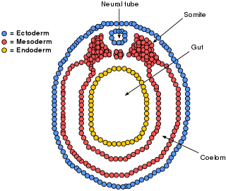
The mesoderm is the middle layer of the three germ layers that develops during gastrulation in the very early development of the embryo of most animals. The outer layer is the ectoderm, and the inner layer is the endoderm.

<span class="mw-page-title-main">Gastrulation</span> Stage in embryonic development in which germ layers form

Gastrulation is the stage in the early embryonic development of most animals, during which the blastula, or in mammals the blastocyst, is reorganized into a two-layered or three-layered embryo known as the gastrula. Before gastrulation, the embryo is a continuous epithelial sheet of cells; by the end of gastrulation, the embryo has begun differentiation to establish distinct cell lineages, set up the basic axes of the body, and internalized one or more cell types including the prospective gut.

<span class="mw-page-title-main">Invagination</span> Process in embryonic development

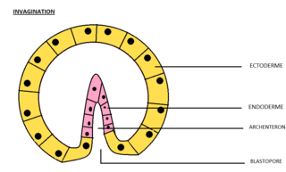
Invagination is the process of a surface folding in on itself to form a cavity, pouch or tube. In developmental biology, invagination is a mechanism that takes place during gastrulation. This mechanism or cell movement happens mostly in the vegetal pole. Invagination consists of the folding of an area of the exterior sheet of cells towards the inside of the blastula. In each organism, the complexity will be different depending on the number of cells. Invagination can be referenced as one of the steps of the establishment of the body plan. The term, originally used in embryology, has been adopted in other disciplines as well.

<span class="mw-page-title-main">Neurulation</span> Embryological process forming the neural tube

Neurulation refers to the folding process in vertebrate embryos, which includes the transformation of the neural plate into the neural tube. The embryo at this stage is termed the neurula.

A germ layer is a primary layer of cells that forms during embryonic development. The three germ layers in vertebrates are particularly pronounced; however, all eumetazoans produce two or three primary germ layers. Some animals, like cnidarians, produce two germ layers making them diploblastic. Other animals such as bilaterians produce a third layer between these two layers, making them triploblastic. Germ layers eventually give rise to all of an animal's tissues and organs through the process of organogenesis.

Organogenesis is the phase of embryonic development that starts at the end of gastrulation and continues until birth. During organogenesis, the three germ layers formed from gastrulation form the internal organs of the organism.

<span class="mw-page-title-main">Animal embryonic development</span> Process by which the embryo forms and develops

In developmental biology, animal embryonic development, also known as animal embryogenesis, is the developmental stage of an animal embryo. Embryonic development starts with the fertilization of an egg cell (ovum) by a sperm cell, (spermatozoon). Once fertilized, the ovum becomes a single diploid cell known as a zygote. The zygote undergoes mitotic divisions with no significant growth and cellular differentiation, leading to development of a multicellular embryo after passing through an organizational checkpoint during mid-embryogenesis. In mammals, the term refers chiefly to the early stages of prenatal development, whereas the terms fetus and fetal development describe later stages.

<span class="mw-page-title-main">Neural plate</span>

The neural plate is a key developmental structure that serves as the basis for the nervous system. Cranial to the primitive node of the embryonic primitive streak, ectodermal tissue thickens and flattens to become the neural plate. The region anterior to the primitive node can be generally referred to as the neural plate. Cells take on a columnar appearance in the process as they continue to lengthen and narrow. The ends of the neural plate, known as the neural folds, push the ends of the plate up and together, folding into the neural tube, a structure critical to brain and spinal cord development. This process as a whole is termed primary neurulation.

<span class="mw-page-title-main">Neurula</span> Embryo at the early stage of development in which neurulation occurs

A neurula is a vertebrate embryo at the early stage of development in which neurulation occurs. The neurula stage is preceded by the gastrula stage; consequentially, neurulation is preceded by gastrulation. Neurulation marks the beginning of the process of organogenesis.

<span class="mw-page-title-main">Neural crest</span> Pluripotent embyronic cell group giving rise to diverse cell lineages

Neural crest cells are a temporary group of cells that arise from the embryonic ectoderm germ layer, and in turn give rise to a diverse cell lineage—including melanocytes, craniofacial cartilage and bone, smooth muscle, peripheral and enteric neurons and glia.

<span class="mw-page-title-main">Neural fold</span> Structure arising during embryonic development of birds and mammals

The neural fold is a structure that arises during neurulation in the embryonic development of both birds and mammals among other organisms. This structure is associated with primary neurulation, meaning that it forms by the coming together of tissue layers, rather than a clustering, and subsequent hollowing out, of individual cells. In humans, the neural folds are responsible for the formation of the anterior end of the neural tube. The neural folds are derived from the neural plate, a preliminary structure consisting of elongated ectoderm cells. The folds give rise to neural crest cells, as well as bringing about the formation of the neural tube.

<span class="mw-page-title-main">Epiblast</span> Embryonic inner cell mass tissue that forms the embryo itself, through the three germ layers

In amniote embryonic development, the epiblast is one of two distinct cell layers arising from the inner cell mass in the mammalian blastocyst, or from the blastula in reptiles and birds, the other layer is the hypoblast. It drives the embryo proper through its differentiation into the three primary germ layers, ectoderm, mesoderm and endoderm, during gastrulation. The amniotic ectoderm and extraembryonic mesoderm also originate from the epiblast.

<span class="mw-page-title-main">Mesenchyme</span> Type of animal embryonic connective tissue

Mesenchyme is a type of loosely organized animal embryonic connective tissue of undifferentiated cells that give rise to most tissues, such as skin, blood or bone. The interactions between mesenchyme and epithelium help to form nearly every organ in the developing embryo.

<span class="mw-page-title-main">Bilaminar embryonic disc</span>

The bilaminar embryonic disc, bilaminar blastoderm or embryonic disc is the distinct two-layered structure of cells formed in an embryo. In the development of the human embryo this takes place by day eight. It is formed when the inner cell mass, also known as the embryoblast, forms a bilaminar disc of two layers, an upper layer called the epiblast and a lower layer called the hypoblast, which will eventually form into fetus. These two layers of cells are stretched between two fluid-filled cavities at either end: the primitive yolk sac and the amniotic sac.

<span class="mw-page-title-main">Eye development</span> Formation of the eye during embryonic development

Eye formation in the human embryo begins at approximately three weeks into embryonic development and continues through the tenth week. Cells from both the mesodermal and the ectodermal tissues contribute to the formation of the eye. Specifically, the eye is derived from the neuroepithelium, surface ectoderm, and the extracellular mesenchyme which consists of both the neural crest and mesoderm.

<span class="mw-page-title-main">Fish development</span>

The development of fishes is unique in some specific aspects compared to the development of other animals.

<span class="mw-page-title-main">Human embryonic development</span> Development and formation of the human embryo

Human embryonic development or human embryogenesis is the development and formation of the human embryo. It is characterised by the processes of cell division and cellular differentiation of the embryo that occurs during the early stages of development. In biological terms, the development of the human body entails growth from a one-celled zygote to an adult human being. Fertilization occurs when the sperm cell successfully enters and fuses with an egg cell (ovum). The genetic material of the sperm and egg then combine to form the single cell zygote and the germinal stage of development commences. Embryonic development in the human, covers the first eight weeks of development; at the beginning of the ninth week the embryo is termed a fetus. The eight weeks has 23 stages.

<span class="mw-page-title-main">Surface ectoderm</span> Embryonic division of the ectoderm

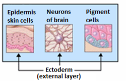
The surface ectoderm, AKA external ectoderm, is one of the two early embryonic divisions of the ectoderm. The other early division of the ectoderm is the neuroectoderm.

<span class="mw-page-title-main">Ectoderm specification</span> Stage in embryonic development

In Xenopus laevis, the specification of the three germ layers occurs at the blastula stage. Great efforts have been made to determine the factors that specify the endoderm and mesoderm. On the other hand, only a few examples of genes that are required for ectoderm specification have been described in the last decade. The first molecule identified to be required for the specification of ectoderm was the ubiquitin ligase Ectodermin ; later, it was found that the deubiquitinating enzyme, FAM/USP9x, is able to overcome the effects of ubiquitination made by Ectodermin in Smad4. Two transcription factors have been proposed to control gene expression of ectodermal specific genes: POU91/Oct3/4 and FoxIe1/Xema. A new factor specific for the ectoderm, XFDL156, has shown to be essential for suppression of mesoderm differentiation from pluripotent cells.

References

  1. Langman's Medical Embryology, 11th edition. 2010.
  2. 1 2 3 4 5 6 7 Gilbert, Scott F. Developmental Biology. 9th ed. Sunderland, MA: Sinauer Associates, 2010: 333-370. Print.
  3. 1 2 "Derivation of Tissues | SEER Training". training.seer.cancer.gov.
  4. 1 2 Marieb, Elaine N.; Hoehn, Katja (2019). Human Anatomy & Physiology. United States of America: Pearson. pp. 146, 482–483, 1102–1106. ISBN   978-0-13-458099-9.
  5. Baer KE von (1986) In: Oppenheimer J (ed.) and Schneider H (transl.), Autobiography of Dr. Karl Ernst von Baer. Canton, MA: Science History Publications.
  6. 1 2 Hosseini, Hadi S.; Garcia, Kara E.; Taber, Larry A. (2017). "A new hypothesis for foregut and heart tube formation based on differential growth and actomyosin contraction". Development. 144 (13): 2381–2391. doi: 10.1242/dev.145193 . PMC   5536863 . PMID   28526751.
  7. O'Rahilly, R; Müller, F (1994). "Neurulation in the Normal Human Embryo". Ciba Foundation Symposium 181 ‐ Neural Tube Defects. Novartis Foundation Symposia. Vol. 181. pp. 70–82. doi:10.1002/9780470514559.ch5. ISBN   9780470514559. PMID   8005032.{{cite book}}: |journal= ignored (help)
  8. Pispa, J; Thesleff, I (Oct 15, 2003). "Mechanisms of ectodermal organogenesis". Developmental Biology. 262 (2): 195–205. doi: 10.1016/S0012-1606(03)00325-7 . PMID   14550785.
  9. 1 2 Tai, Y. Y.; Chen, R. S.; Lin, Y.; Ling, T. Y.; Chen, M. H. (2012). "FGF-9 accelerates epithelial invagination for ectodermal organogenesis in real time bioengineered organ manipulation". Cell Communication and Signaling. 10 (1): 34. doi: 10.1186/1478-811X-10-34 . PMC   3515343 . PMID   23176204.
  10. Priolo, M.; Laganà, C (September 2001). "Ectodermal Dysplasias: A New Clinical-Genetic Classification". Journal of Medical Genetics. 38 (9): 579–585. doi:10.1136/jmg.38.9.579. PMC   1734928 . PMID   11546825.
  11. Clarke, A.; Phillips, D. I.; Brown, R.; Harper, P. S. (1987). "Clinical Aspects of X-linked Hypohidrotic Ectodermal Dysplasia". Archives of Disease in Childhood. 62 (10): 989–96. doi:10.1136/adc.62.10.989. PMC   1778691 . PMID   2445301.
  12. Bayes, M.; Hartung, A. J.; Ezer, S.; Pispa, J.; Thesleff, I.; Srivastava, A. K.; Kere, J. (1998). "The Anhidrotic Ectodermal Dysplasia Gene (EDA) Undergoes Alternative Splicing and Encodes Ectodysplasin-A with Deletion Mutations in Collagenous Repeats". Human Molecular Genetics. 7 (11): 1661–1669. doi: 10.1093/hmg/7.11.1661 . PMID   9736768.