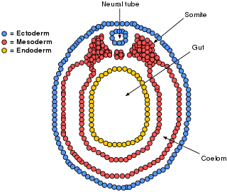
The mesoderm is the middle layer of the three germ layers that develops during gastrulation in the very early development of the embryo of most animals. The outer layer is the ectoderm, and the inner layer is the endoderm.

Gastrulation is the stage in the early embryonic development of most animals, during which the blastula, or in mammals the blastocyst, is reorganized into a two-layered or three-layered embryo known as the gastrula. Before gastrulation, the embryo is a continuous epithelial sheet of cells; by the end of gastrulation, the embryo has begun differentiation to establish distinct cell lineages, set up the basic axes of the body, and internalized one or more cell types including the prospective gut.

The ectoderm is one of the three primary germ layers formed in early embryonic development. It is the outermost layer, and is superficial to the mesoderm and endoderm. It emerges and originates from the outer layer of germ cells. The word ectoderm comes from the Greek ektos meaning "outside", and derma meaning "skin".

Endoderm is the innermost of the three primary germ layers in the very early embryo. The other two layers are the ectoderm and mesoderm. Cells migrating inward along the archenteron form the inner layer of the gastrula, which develops into the endoderm.

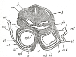
The somites are a set of bilaterally paired blocks of paraxial mesoderm that form in the embryonic stage of somitogenesis, along the head-to-tail axis in segmented animals. In vertebrates, somites subdivide into the dermatomes, myotomes, sclerotomes and syndetomes that give rise to the vertebrae of the vertebral column, rib cage, part of the occipital bone, skeletal muscle, cartilage, tendons, and skin.

The yolk sac is a membranous sac attached to an embryo, formed by cells of the hypoblast layer of the bilaminar embryonic disc. This is alternatively called the umbilical vesicle by the Terminologia Embryologica (TE), though yolk sac is far more widely used. In humans, the yolk sac is important in early embryonic blood supply, and much of it is incorporated into the primordial gut during the fourth week of embryonic development.
The archenteron, also called the gastrocoel, the primitive digestive tube or the primitive gut, is the internal cavity of the primitive gastrointestinal tract that forms during gastrulation in a developing animal embryo. It develops into the endoderm and mesoderm of the animal.

The pharyngeal arches, also known as visceral arches, are structures seen in the embryonic development of vertebrates that are recognisable precursors for many structures. In fish, the arches are known as the branchial arches, or gill arches.
In the embryonic development of vertebrates, pharyngeal pouches form on the endodermal side between the pharyngeal arches. The pharyngeal grooves form the lateral ectodermal surface of the neck region to separate the arches.
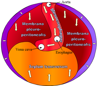
The septum transversum is a thick mass of cranial mesenchyme, formed in the embryo, that gives rise to parts of the thoracic diaphragm and the ventral mesentery of the foregut in the developed human being and other mammals.
The lateral plate mesoderm is the mesoderm that is found at the periphery of the embryo. It is to the side of the paraxial mesoderm, and further to the axial mesoderm. The lateral plate mesoderm is separated from the paraxial mesoderm by a narrow region of intermediate mesoderm. The mesoderm is the middle layer of the three germ layers, between the outer ectoderm and inner endoderm.

The eyes begin to develop as a pair of diverticula (pouches) from the lateral aspects of the forebrain. These diverticula make their appearance before the closure of the anterior end of the neural tube; after the closure of the tube around the 4th week of development, they are known as the optic vesicles. Previous studies of optic vesicles suggest that the surrounding extraocular tissues – the surface ectoderm and extraocular mesenchyme – are necessary for normal eye growth and differentiation.

The cloacal membrane is the membrane that covers the embryonic cloaca during the development of the urinary and reproductive organs.

In the anatomy of an embryo, the somatopleure is a structure created during embryogenesis when the lateral plate mesoderm splits into two layers. The outer layer becomes applied to the inner surface of the ectoderm, and with it (partially) forms the somatopleure.

Mesenchyme is a type of loosely organized animal embryonic connective tissue of undifferentiated cells that give rise to most tissues, such as skin, blood or bone. The interactions between mesenchyme and epithelium help to form nearly every organ in the developing embryo.

The lung bud sometimes referred to as the respiratory bud forms from the respiratory diverticulum, an embryological endodermal structure that develops into the respiratory tract organs such as the larynx, trachea, bronchi and lungs. It arises from part of the laryngotracheal tube.

Human embryonic development or human embryogenesis is the development and formation of the human embryo. It is characterised by the processes of cell division and cellular differentiation of the embryo that occurs during the early stages of development. In biological terms, the development of the human body entails growth from a one-celled zygote to an adult human being. Fertilization occurs when the sperm cell successfully enters and fuses with an egg cell (ovum). The genetic material of the sperm and egg then combine to form the single cell zygote and the germinal stage of development commences. Embryonic development in the human, covers the first eight weeks of development; at the beginning of the ninth week the embryo is termed a fetus. The eight weeks have 23 stages.

In amniote embryology, the hypoblast is one of two distinct layers arising from the inner cell mass in the mammalian blastocyst, or from the blastodisc in reptiles and birds. The hypoblast gives rise to the yolk sac, which in turn gives rise to the chorion.
In the development of vertebrate animals, the prechordal plate is a "uniquely thickened portion" of the endoderm that is in contact with ectoderm immediately rostral to the cephalic tip of the notochord. It is the most likely origin of the rostral cranial mesoderm.
The development of the digestive system in the human embryo concerns the epithelium of the digestive system and the parenchyma of its derivatives, which originate from the endoderm. Connective tissue, muscular components, and peritoneal components originate in the mesoderm. Different regions of the gut tube such as the esophagus, stomach, duodenum, etc. are specified by a retinoic acid gradient that causes transcription factors unique to each region to be expressed. Differentiation of the gut and its derivatives depends upon reciprocal interactions between the gut endoderm and its surrounding mesoderm. Hox genes in the mesoderm are induced by a Hedgehog signaling pathway secreted by gut endoderm and regulate the craniocaudal organization of the gut and its derivatives. The gut system extends from the oropharyngeal membrane to the cloacal membrane and is divided into the foregut, midgut, and hindgut.