Organogenesis

Last updated

Organogenesis is the phase of embryonic development that starts at the end of gastrulation and continues until birth. During organogenesis, the three germ layers formed from gastrulation (the ectoderm, endoderm, and mesoderm) form the internal organs of the organism. [1]

Contents

The endoderm of vertebrates produces tissue within the lungs, thyroid, and pancreas. The mesoderm aids in the production of cardiac muscle, skeletal muscle, smooth muscle, tissues within the kidneys, and red blood cells. The ectoderm produces tissues within the epidermis and aids in the formation of neurons within the brain, and melanocytes. Germ layers.jpg
The endoderm of vertebrates produces tissue within the lungs, thyroid, and pancreas. The mesoderm aids in the production of cardiac muscle, skeletal muscle, smooth muscle, tissues within the kidneys, and red blood cells. The ectoderm produces tissues within the epidermis and aids in the formation of neurons within the brain, and melanocytes.

The cells of each of the three germ layers undergo differentiation, a process where less-specialized cells become more-specialized through the expression of a specific set of genes. Cell differentiation is driven by cell signaling cascades. [2] Differentiation is influenced by extracellular signals such as growth factors that are exchanged to adjacent cells which is called juxtracrine signaling or to neighboring cells over short distances which is called paracrine signaling. [3] Intracellular signals – a cell signaling itself (autocrine signaling) – also play a role in organ formation. These signaling pathways allow for cell rearrangement and ensure that organs form at specific sites within the organism. [1] The organogenesis process can be studied using embryos and organoids. [4]

Organs produced by the germ layers

Neural precursor cells fold and elongate to form the neural tube. Mesoderm cells condense to form a rod which will send out signals to redirect the ectoderm cells above. This fold along the neural tube sets up the vertebrate central nervous system. Development of the neural tube.png
Neural precursor cells fold and elongate to form the neural tube. Mesoderm cells condense to form a rod which will send out signals to redirect the ectoderm cells above. This fold along the neural tube sets up the vertebrate central nervous system.

The endoderm is the inner most germ layer of the embryo which gives rise to gastrointestinal and respiratory organs by forming epithelial linings and organs such as the liver, lungs, and pancreas. [5] The mesoderm or middle germ layer of the embryo will form the blood, heart, kidney, muscles, and connective tissues. [5] The ectoderm or outermost germ layer of the developing embryo forms epidermis, the brain, and the nervous system. [5]

Mechanism of organ formation

While each germ layer forms specific organs, in the 1820s, embryologist Heinz Christian Pander discovered that the germ layers cannot form their respective organs without the cellular interactions from other tissues. [1] In humans, internal organs begin to develop within 3–8 weeks after fertilization. The germ layers form organs by three processes: folds, splits, and condensation. [6] Folds form in the germinal sheet of cells and usually form an enclosed tube which you can see in the development of vertebrates neural tube. Splits or pockets may form in the germinal sheet of cells forming vesicles or elongations. The lungs and glands of the organism may develop this way. [6]

A primary step in organogenesis for chordates is the development of the notochord, which induces the formation of the neural plate, and ultimately the neural tube in vertebrate development. The development of the neural tube will give rise to the brain and spinal cord. [1] Vertebrates develop a neural crest that differentiates into many structures, including bones, muscles, and components of the central nervous system. Differentiation of the ectoderm into the neural crest, neural tube, and surface ectoderm is sometimes referred to as neurulation and the embryo in this phase is the neurula. The coelom of the body forms from a split of the mesoderm along the somite axis [1]

Plant organogenesis

In plants, organogenesis occurs continuously and only stops when the plant dies. In the shoot, the shoot apical meristems regularly produce new lateral organs (leaves or flowers) and lateral branches. In the root, new lateral roots form from weakly differentiated internal tissue (e.g. the xylem-pole pericycle in the model plant Arabidopsis thaliana ). In vitro and in response to specific cocktails of hormones (mainly auxins and cytokinins), most plant tissues can de-differentiate and form a mass of dividing totipotent stem cells called a callus. Organogenesis can then occur from those cells. The type of organ that is formed depends on the relative concentrations of the hormones in the medium. Plant organogenesis can be induced in tissue culture and used to regenerate plants. [7]

See also

Related Research Articles

Developmental biology is the study of the process by which animals and plants grow and develop. Developmental biology also encompasses the biology of regeneration, asexual reproduction, metamorphosis, and the growth and differentiation of stem cells in the adult organism.

<span class="mw-page-title-main">Ontogeny</span> Origination and development of an organism

Ontogeny is the origination and development of an organism, usually from the time of fertilization of the egg to adult. The term can also be used to refer to the study of the entirety of an organism's lifespan.

<span class="mw-page-title-main">Embryo</span> Multicellular diploid eukaryote in its earliest stage of development

An embryo is an initial stage of development of a multicellular organism. In organisms that reproduce sexually, embryonic development is the part of the life cycle that begins just after fertilization of the female egg cell by the male sperm cell. The resulting fusion of these two cells produces a single-celled zygote that undergoes many cell divisions that produce cells known as blastomeres. The blastomeres are arranged as a solid ball that when reaching a certain size, called a morula, takes in fluid to create a cavity called a blastocoel. The structure is then termed a blastula, or a blastocyst in mammals.

<span class="mw-page-title-main">Mesoderm</span> Middle germ layer of embryonic development

The mesoderm is the middle layer of the three germ layers that develops during gastrulation in the very early development of the embryo of most animals. The outer layer is the ectoderm, and the inner layer is the endoderm.
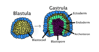
<span class="mw-page-title-main">Gastrulation</span> Stage in embryonic development in which germ layers form

Gastrulation is the stage in the early embryonic development of most animals, during which the blastula, or in mammals the blastocyst is reorganized into a two-layered or three-layered embryo known as the gastrula. Before gastrulation, the embryo is a continuous epithelial sheet of cells; by the end of gastrulation, the embryo has begun differentiation to establish distinct cell lineages, set up the basic axes of the body, and internalized one or more cell types including the prospective gut.

<span class="mw-page-title-main">Ectoderm</span> Outer germ layer of embryonic development

The ectoderm is one of the three primary germ layers formed in early embryonic development. It is the outermost layer, and is superficial to the mesoderm and endoderm. It emerges and originates from the outer layer of germ cells. The word ectoderm comes from the Greek ektos meaning "outside", and derma meaning "skin".

<span class="mw-page-title-main">Endoderm</span> Inner germ layer of embryonic development

Endoderm is the innermost of the three primary germ layers in the very early embryo. The other two layers are the ectoderm and mesoderm. Cells migrating inward along the archenteron form the inner layer of the gastrula, which develops into the endoderm.
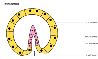
<span class="mw-page-title-main">Invagination</span> Process in embryonic development

Invagination is the process of a surface folding in on itself to form a cavity, pouch or tube. In developmental biology, invagination is a mechanism that takes place during gastrulation. This mechanism or cell movement happens mostly in the vegetal pole. Invagination consists of the folding of an area of the exterior sheet of cells towards the inside of the blastula. In each organism, the complexity will be different depending on the number of cells. Invagination can be referenced as one of the steps of the establishment of the body plan. The term, originally used in embryology, has been adopted in other disciplines as well.

<span class="mw-page-title-main">Neurulation</span> Embryological process forming the neural tube

Neurulation refers to the folding process in vertebrate embryos, which includes the transformation of the neural plate into the neural tube. The embryo at this stage is termed the neurula.

A germ layer is a primary layer of cells that forms during embryonic development. The three germ layers in vertebrates are particularly pronounced; however, all eumetazoans produce two or three primary germ layers. Some animals, like cnidarians, produce two germ layers making them diploblastic. Other animals such as bilaterians produce a third layer between these two layers, making them triploblastic. Germ layers eventually give rise to all of an animal's tissues and organs through the process of organogenesis.

<span class="mw-page-title-main">Animal embryonic development</span> Process by which the embryo forms and develops

In developmental biology, animal embryonic development, also known as animal embryogenesis, is the developmental stage of an animal embryo. Embryonic development starts with the fertilization of an egg cell (ovum) by a sperm cell, (spermatozoon). Once fertilized, the ovum becomes a single diploid cell known as a zygote. The zygote undergoes mitotic divisions with no significant growth and cellular differentiation, leading to development of a multicellular embryo after passing through an organizational checkpoint during mid-embryogenesis. In mammals, the term refers chiefly to the early stages of prenatal development, whereas the terms fetus and fetal development describe later stages.
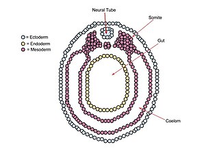
<span class="mw-page-title-main">Neurula</span> Embryo at the early stage of development in which neurulation occurs

A neurula is a vertebrate embryo at the early stage of development in which neurulation occurs. The neurula stage is preceded by the gastrula stage; consequentially, neurulation is preceded by gastrulation. Neurulation marks the beginning of the process of organogenesis.

Histogenesis is the formation of different tissues from undifferentiated cells. These cells are constituents of three primary germ layers, the endoderm, mesoderm, and ectoderm. The science of the microscopic structures of the tissues formed within histogenesis is termed histology.

The primitive node is the organizer for gastrulation in most amniote embryos. In birds it is known as Hensen's node, and in amphibians it is known as the Spemann-Mangold organizer. It is induced by the Nieuwkoop center in amphibians, or by the posterior marginal zone in amniotes including birds.

<span class="mw-page-title-main">Epiblast</span> Embryonic inner cell mass tissue that forms the embryo itself, through the three germ layers

In amniote embryonic development, the epiblast is one of two distinct cell layers arising from the inner cell mass in the mammalian blastocyst, or from the blastula in reptiles and birds, the other layer is the hypoblast. It drives the embryo proper through its differentiation into the three primary germ layers, ectoderm, mesoderm and endoderm, during gastrulation. The amniotic ectoderm and extraembryonic mesoderm also originate from the epiblast.
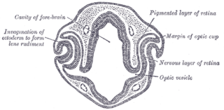
<span class="mw-page-title-main">Eye development</span> Formation of the eye during embryonic development

Eye formation in the human embryo begins at approximately three weeks into embryonic development and continues through the tenth week. Cells from both the mesodermal and the ectodermal tissues contribute to the formation of the eye. Specifically, the eye is derived from the neuroepithelium, surface ectoderm, and the extracellular mesenchyme which consists of both the neural crest and mesoderm.

Convergent extension (CE), sometimes called convergence and extension (C&E), is the process by which the tissue of an embryo is restructured to converge (narrow) along one axis and extend (elongate) along a perpendicular axis by cellular movement.

<span class="mw-page-title-main">Fish development</span>

The development of fishes is unique in some specific aspects compared to the development of other animals.

<span class="mw-page-title-main">Human embryonic development</span> Development and formation of the human embryo

Human embryonic development, or human embryogenesis, is the development and formation of the human embryo. It is characterised by the processes of cell division and cellular differentiation of the embryo that occurs during the early stages of development. In biological terms, the development of the human body entails growth from a one-celled zygote to an adult human being. Fertilization occurs when the sperm cell successfully enters and fuses with an egg cell (ovum). The genetic material of the sperm and egg then combine to form the single cell zygote and the germinal stage of development commences. Embryonic development in the human, covers the first eight weeks of development; at the beginning of the ninth week the embryo is termed a fetus. The eight weeks has 23 stages.

<span class="mw-page-title-main">Hypoblast</span> Embryonic inner cell mass tissue that forms the yolk sac and, later, chorion

In amniote embryology, the hypoblast is one of two distinct layers arising from the inner cell mass in the mammalian blastocyst, or from the blastodisc in reptiles and birds. The hypoblast gives rise to the yolk sac, which in turn gives rise to the chorion.

References

  1. 1 2 3 4 5 Gilbert, S. F.; Barresi, M. J. F. (2017-05-01). "Developmental Biology, 11Th Edition 2016". American Journal of Medical Genetics Part A. 173 (5): 1430. doi:10.1002/ajmg.a.38166. ISSN   1552-4833.
  2. Rankin, Scott (2018). "Timing is everything: Reiterative Wnt, BMP and RA signaling regulate developmental competence during endoderm organogenesis". Developmental Biology. 434 (1): 121–132. doi:10.1016/j.ydbio.2017.11.018. PMC   5785443 . PMID   29217200 via NCBI.
  3. Edlund, Helena (July 2002). "Organogenesis: Pancreatic organogenesis — developmental mechanisms and implications for therapy". Nature Reviews Genetics. 3 (7): 524–532. doi:10.1038/nrg841. ISSN   1471-0064. PMID   12094230. S2CID   2436869.
  4. Ader, Marius; Tanaka, Elly M (2014). "Modeling human development in 3D culture". Current Opinion in Cell Biology. 31: 23–28. doi:10.1016/j.ceb.2014.06.013. PMID   25033469.
  5. 1 2 3 Kiecker, Clemens; Bates, Thomas; Bell, Esther (2016-03-01). "Molecular specification of germ layers in vertebrate embryos". Cellular and Molecular Life Sciences. 73 (5): 923–947. doi:10.1007/s00018-015-2092-y. ISSN   1420-682X. PMC   4744249 . PMID   26667903.
  6. 1 2 "Animal development – Embryonic induction". Encyclopedia Britannica. Retrieved 2018-04-04.
  7. "Plant and Soil Sciences eLibrary". passel.unl.edu. Retrieved 2018-04-04.