Related Research Articles

The vagus nerve, also known as the tenth cranial nerve, cranial nerve X, or simply CN X, is a cranial nerve that carries sensory fibers that create a pathway that interfaces with the parasympathetic control of the heart, lungs, and digestive tract. It comprises two nerves—the left and right vagus nerves—but they are typically referred to collectively as a single subsystem.

The autonomic nervous system (ANS), formerly referred to as the vegetative nervous system, is a division of the nervous system that operates internal organs, smooth muscle and glands. The autonomic nervous system is a control system that acts largely unconsciously and regulates bodily functions, such as the heart rate, its force of contraction, digestion, respiratory rate, pupillary response, urination, and sexual arousal. This system is the primary mechanism in control of the fight-or-flight response.

The splanchnic nerves are paired visceral nerves, carrying fibers of the autonomic nervous system as well as sensory fibers from the organs. All carry sympathetic fibers except for the pelvic splanchnic nerves, which carry parasympathetic fibers.

The levator ani is a broad, thin muscle group, situated on either side of the pelvis. It is formed from three muscle components: the pubococcygeus, the iliococcygeus, and the puborectalis.
The parasympathetic nervous system (PSNS) is one of the three divisions of the autonomic nervous system, the others being the sympathetic nervous system and the enteric nervous system. The enteric nervous system is sometimes considered part of the autonomic nervous system, and sometimes considered an independent system.

A spinal nerve is a mixed nerve, which carries motor, sensory, and autonomic signals between the spinal cord and the body. In the human body there are 31 pairs of spinal nerves, one on each side of the vertebral column. These are grouped into the corresponding cervical, thoracic, lumbar, sacral and coccygeal regions of the spine. There are eight pairs of cervical nerves, twelve pairs of thoracic nerves, five pairs of lumbar nerves, five pairs of sacral nerves, and one pair of coccygeal nerves. The spinal nerves are part of the peripheral nervous system.
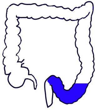
The sigmoid colon is the part of the large intestine that is closest to the rectum and anus. It forms a loop that averages about 35–40 centimetres (14–16 in) in length. The loop is typically shaped like a Greek letter sigma (ς) or Latin letter S. This part of the colon normally lies within the pelvis, but due to its freedom of movement it is liable to be displaced into the abdominal cavity.

A dermatome is an area of skin that is mainly supplied by afferent nerve fibres from the dorsal root of any given spinal nerve. There are 8 cervical nerves , 12 thoracic nerves, 5 lumbar nerves and 5 sacral nerves. Each of these nerves relays sensation from a particular region of skin to the brain.

The internal anal sphincter, IAS, or sphincter ani internus is a ring of smooth muscle that surrounds about 2.5–4.0 cm of the anal canal. It is about 5 mm thick, and is formed by an aggregation of the smooth (involuntary) circular muscle fibers of the rectum. it terminates distally about 6 mm from the anal orifice.

A nerve plexus is a plexus of intersecting nerves. A nerve plexus is composed of afferent and efferent fibers that arise from the merging of the anterior rami of spinal nerves and blood vessels. There are five spinal nerve plexuses, except in the thoracic region, as well as other forms of autonomic plexuses, many of which are a part of the enteric nervous system. The nerves that arise from the plexuses have both sensory and motor functions. These functions include muscle contraction, the maintenance of body coordination and control, and the reaction to sensations such as heat, cold, pain, and pressure. There are several plexuses in the body, including:

The superior hypogastric plexus is a plexus of nerves situated on the vertebral bodies anterior to the bifurcation of the abdominal aorta. It bifurcates to form the left and the right hypogastric nerve. The SHP is the continuation of the abdominal aortic plexus.
The sympathetic ganglia, or paravertebral ganglia, are autonomic ganglia of the sympathetic nervous system. Ganglia are 20,000 to 30,000 afferent and efferent nerve cell bodies that run along on either side of the spinal cord. Afferent nerve cell bodies bring information from the body to the brain and spinal cord, while efferent nerve cell bodies bring information from the brain and spinal cord to the rest of the body. The cell bodies create long sympathetic chains that are on either side of the spinal cord. They also form para- or pre-vertebral ganglia of gross anatomy.

The pelvic cavity is a body cavity that is bounded by the bones of the pelvis. Its oblique roof is the pelvic inlet. Its lower boundary is the pelvic floor.

The lateral grey column is one of the three grey columns of the spinal cord ; the others being the anterior and posterior grey columns. The lateral grey column is primarily involved with activity in the sympathetic division of the autonomic motor system. It projects to the side as a triangular field in the thoracic and upper lumbar regions of the postero-lateral part of the anterior grey column.
The inferior hypogastric plexus is a network of nerves that supplies the organs of the pelvic cavity. The inferior hypogastric plexus gives rise to the prostatic plexus in males and the uterovaginal plexus in females.

Sacral splanchnic nerves are splanchnic nerves that connect the inferior hypogastric plexus to the sympathetic trunk in the pelvis.

The hypogastric nerves are the continuation of the superior hypogastric plexus that descend into the pelvis anterior the sacrum and become the inferior hypogastric plexuses on either side of pelvic organs. The hypogastric nerves serve as a pathway for autonomic fibers to communicate between the lower abdomen and pelvis.

The general visceral afferent (GVA) fibers conduct sensory impulses from the internal organs, glands, and blood vessels to the central nervous system. They are considered to be part of the visceral nervous system, which is closely related to the autonomic nervous system, but 'visceral nervous system' and 'autonomic nervous system' are not direct synonyms and care should be taken when using these terms. Unlike the efferent fibers of the autonomic nervous system, the afferent fibers are not classified as either sympathetic or parasympathetic.
The lumbar ganglia are paravertebral ganglia located in the inferior portion of the sympathetic trunk. The lumbar portion of the sympathetic trunk typically has 4 lumbar ganglia. The lumbar splanchnic nerves arise from the ganglia here, and contribute sympathetic efferent fibers to the nearby plexuses. The first two lumbar ganglia have both white and gray rami communicates.

The classification of peripheral nerves in the peripheral nervous system (PNS) groups the nerves into two main groups, the somatic and the autonomic nervous systems. Together, these two systems provide information regarding the location and status of the limbs, organs, and the remainder of the body to the central nervous system (CNS) via nerves and ganglia present outside of the spinal cord and brain. The somatic nervous system directs all voluntary movements of the skeletal muscles, and can be sub-divided into afferent and efferent neuronal flow. The autonomic nervous system is divided primarily into the sympathetic and parasympathetic nervous systems with a third system, the enteric nervous system, receiving less recognition.
References
- 1 2 3 Drake, Richard L.; Vogl, Wayne; Tibbitts, Adam W.M. Mitchell; illustrations by Richard; Richardson, Paul (2005). Gray's anatomy for students. Philadelphia: Elsevier/Churchill Livingstone. p. 423. ISBN 978-0-8089-2306-0.
- 1 2 3 4 5 Oakes, W. Jerry; Rocque, Brandon G. (2015-01-01), Tubbs, R. Shane; Rizk, Elias; Shoja, Mohammadali M.; Loukas, Marios (eds.), "Chapter 25 - Dorsal Rhizotomy for Spasticity", Nerves and Nerve Injuries, San Diego: Academic Press, pp. 383–391, ISBN 978-0-12-802653-3 , retrieved 2021-01-06
- 1 2 3 4 Bharucha, ADIL E.; Klingele, CHRISTOPHER J. (2005-01-01), Dyck, Peter J.; Thomas, P. K. (eds.), "Chapter 13 - Autonomic and Somatic Systems to the Anorectum and Pelvic Floor", Peripheral Neuropathy (Fourth Edition), Philadelphia: W.B. Saunders, pp. 279–298, ISBN 978-0-7216-9491-7 , retrieved 2021-01-06