
The parasympathetic nervous system (PSNS) is one of the three divisions of the autonomic nervous system, the others being the sympathetic nervous system and the enteric nervous system. The enteric nervous system is sometimes considered part of the autonomic nervous system, and sometimes considered an independent system.

The corpus spongiosum is the mass of spongy tissue surrounding the male urethra within the penis. It is also called the corpus cavernosum urethrae in older texts.

A fascia is a generic term for macroscopic membranous bodily structures. Fasciae are classified as superficial, visceral or deep, and further designated according to their anatomical location.

The omohyoid muscle is a muscle in the neck. It is one of the infrahyoid muscles. It consists of two bellies separated by an intermediate tendon. Its inferior belly is attached to the scapula; its superior belly is attached to the hyoid bone. Its intermediate tendon is anchored to the clavicle and first rib by a fascial sling. The omohyoid is innervated by the ansa cervicalis of the cervical plexus. It acts to depress the hyoid bone.

In human anatomy, the sacral plexus is a nerve plexus which provides motor and sensory nerves for the posterior thigh, most of the lower leg and foot, and part of the pelvis. It is part of the lumbosacral plexus and emerges from the lumbar vertebrae and sacral vertebrae (L4-S4). A sacral plexopathy is a disorder affecting the nerves of the sacral plexus, usually caused by trauma, nerve compression, vascular disease, or infection. Symptoms may include pain, loss of motor control, and sensory deficits.

A corpus cavernosum penis (singular) is one of a pair of sponge-like regions of erectile tissue, which contain most of the blood in the penis during an erection.

The corpus cavernosum of clitoris is one of a pair of sponge-like regions of erectile tissue of the clitoris. It is made of a sponge-like tissue that fills with blood during erection. This is homologous to the corpus cavernosum penis. The term corpora cavernosa literally means "cave-like bodies".

The inferior hypogastric plexus is a network of nerves that supplies the organs of the pelvic cavity. The inferior hypogastric plexus gives rise to the prostatic plexus in males and the uterovaginal plexus in females.
In human male anatomy, the dorsal veins of the penis are blood vessels that drain the shaft, the skin and the glans of the human penis. They are typically located in the midline on the dorsal aspect of the penis and they comprise the superficial dorsal veinof the penis, that lies in the subcutaneous tissue of the shaft, and the deep dorsal veinof the penis, that lies beneath the deep fascia.
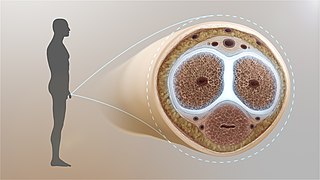
The tunica albuginea is the fibrous envelope that extends the length of the corpora cavernosa penis and corpus spongiosum penis. It is a bi-layered structure that includes an outer longitudinal layer and an inner circular layer.

The prostatic veins form a well-marked prostatic plexus which lies partly in the fascial sheath of the prostate and partly between the sheath and the prostatic capsule. It collects blood from the prostate, and the corpora cavernosa of penis. It communicates with the pudendal and vesical plexuses.
The vesical venous plexus is a venous plexus situated at the fundus of the urinary bladder. It collects venous blood from the urinary bladder in both sexes, from the accessory sex glands in males, and from the corpora cavernosa of clitoris in females. It drains into the internal iliac veins via several vesical veins.
The fibrous envelope of the corpus cavernosum urethrae is thinner, whiter in color, and more elastic than that of the corpora cavernosa penis. It is called the trabeculae of corpus spongiosum of penis.
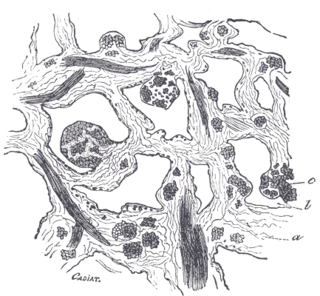
From the internal surface of the fibrous envelope of the corpora cavernosa penis, as well as from the sides of the septum, numerous bands or cords are given off, which cross the interior of these corpora cavernosa in all directions, subdividing them into a number of separate compartments, and giving the entire structure a spongy appearance.

The two crura of penis constitute the root of penis along with the bulb of penis. The two crura flank the bulb - one to each side of the bulb. Each crus is attached at the angle between the perineal membrane and ischiopubic ramus. The deep artery of the penis enters the anterior portion of the crus. Distally, each crus transitions into either corpus spongiosum of the body of the penis.

The helicine arteries of penis are arteries in the penis. They are found in the corpora cavernosa penis.
The cavernous nerves are post-ganglionic parasympathetic nerves that facilitate penile erection and clitoral erection. They arise from cell bodies in the inferior hypogastric plexus where they receive the pre-ganglionic pelvic splanchnic nerves (S2-S4).

In human male anatomy, the root of the penis is the internal and most proximal portion of the human penis that lies in the perineum. Unlike the pendulous body of the penis, which is suspended from the pubic symphysis, the root is attached to the pubic arch of the pelvis and is not visible externally. It is triradiate in form, consisting of three masses of erectile tissue; the two diverging crura, one on either side, and the median bulb of the penis or urethral bulb. Approximately one third of the penis is embedded in the pelvis and can be felt through the scrotum and in the perineum.
In human male anatomy, the septum of the penis or penile septum refers to the fibrous junction (septum) between the two corpora cavernosa of the human penis. The tunica albuginea of the penis forms a thick fibrous coat to the spongy tissue of the corpora cavernosa and corpus spongiosum. The two corpora cavernosa are surrounded by a strong fibrous envelope consisting of superficial and deep fibers. The superficial or outer fibers are longitudinal in direction, and form a single tube which encloses both corpora; the deep or inner fibers are arranged circularly around each corpus and meet in the center. By their junction in the median plane, the inner fibers form the intercavernous septum of the penis.

An erection is a physiological phenomenon in which the penis becomes firm, engorged, and enlarged. Penile erection is the result of a complex interaction of psychological, neural, vascular, and endocrine factors, and is often associated with sexual arousal, sexual attraction or libido, although erections can also be spontaneous. The shape, angle, and direction of an erection vary considerably between humans.
