| Prevertebral plexus | |
|---|---|
 | |
| Anatomical terms of neuroanatomy |
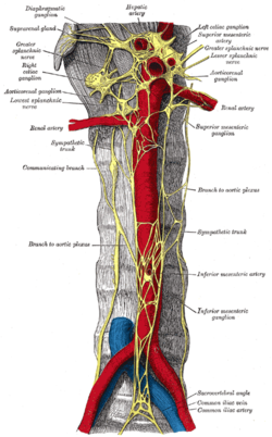
A prevertebral plexus is a nerve plexus which branches from a prevertebral ganglion.
| Prevertebral plexus | |
|---|---|
| | |
| Anatomical terms of neuroanatomy |
A prevertebral plexus is a nerve plexus which branches from a prevertebral ganglion.

The autonomic nervous system (ANS), sometimes called the visceral nervous system and formerly the vegetative nervous system, is a division of the nervous system that operates internal organs, smooth muscle and glands. The autonomic nervous system is a control system that acts largely unconsciously and regulates bodily functions, such as the heart rate, its force of contraction, digestion, respiratory rate, pupillary response, urination, and sexual arousal. This system is the primary mechanism in control of the fight-or-flight response.

The enteric nervous system (ENS) or intrinsic nervous system is one of the three main divisions of the autonomic nervous system (ANS), the other being the sympathetic (SNS) and parasympathetic nervous system (PSNS), and consists of a mesh-like system of neurons that governs the function of the gastrointestinal tract. It is capable of acting independently of the SNS and PSNS, although it may be influenced by them. The ENS is nicknamed the "second brain". It is derived from neural crest cells.

The phrenic nerve is a mixed motor/sensory nerve that originates from the C3-C5 spinal nerves in the neck. The nerve is important for breathing because it provides exclusive motor control of the diaphragm, the primary muscle of respiration. In humans, the right and left phrenic nerves are primarily supplied by the C4 spinal nerve, but there is also a contribution from the C3 and C5 spinal nerves. From its origin in the neck, the nerve travels downward into the chest to pass between the heart and lungs towards the diaphragm.

The cervical plexus is a nerve plexus of the anterior rami of the first four cervical spinal nerves C1-C4. The cervical plexus provides motor innervation to some muscles of the neck, and the diaphragm; it provides sensory innervation to parts of the head, neck, and chest.

The superior thoracic aperture, also known as the thoracic outlet, or thoracic inlet refers to the opening at the top of the thoracic cavity. It is also clinically referred to as the thoracic outlet, in the case of thoracic outlet syndrome. A lower thoracic opening is the inferior thoracic aperture.

In human anatomy, the sacral plexus is a nerve plexus which provides motor and sensory nerves for the posterior thigh, most of the lower leg and foot, and part of the pelvis. It is part of the lumbosacral plexus and emerges from the lumbar vertebrae and sacral vertebrae (L4-S4). A sacral plexopathy is a disorder affecting the nerves of the sacral plexus, usually caused by trauma, nerve compression, vascular disease, or infection. Symptoms may include pain, loss of motor control, and sensory deficits.

The carotid sheath is a condensation of the deep cervical fascia enveloping multiple vital neurovascular structures of the neck, including the common and internal carotid arteries, the internal jugular vein, the vagus nerve, and ansa cervicalis. The carotid sheath helps protects the structures contained therein.

Prevertebral ganglia are sympathetic ganglia situated along the midline, anterior to the aorta and the vertebral column. The prevertebral ganglia are the celiac ganglia, the superior mesenteric ganglion, and the inferior mesenteric ganglion.
The sympathetic trunks are a paired bundle of nerve fibers that run from the base of the skull to the coccyx. They are a major component of the sympathetic nervous system.

The midgut is the portion of the human embryo from which most of the intestines develop. After it bends around the superior mesenteric artery, it is called the "midgut loop". It comprises the portion of the alimentary canal from the end of the foregut at the opening of the bile duct to the hindgut, about two-thirds of the way through the transverse colon.
Each spinal nerve receives a branch called a gray ramus communicans from the adjacent paravertebral ganglion of the sympathetic trunk. The gray rami communicantes contain postganglionic nerve fibers of the sympathetic nervous system and are composed of largely unmyelinated neurons. This is in contrast to the white rami communicantes, in which heavily myelinated neurons give the rami their white appearance.

The celiac ganglia or coeliac ganglia are two large irregularly shaped masses of nerve tissue in the upper abdomen. Part of the sympathetic subdivision of the autonomic nervous system (ANS), the two celiac ganglia are the largest ganglia in the ANS, and they innervate most of the digestive tract.

The sympathetic ganglia, or paravertebral ganglia, are autonomic ganglia of the sympathetic nervous system. Ganglia are 20,000 to 30,000 afferent and efferent nerve cell bodies that run along on either side of the spinal cord. Afferent nerve cell bodies bring information from the body to the brain and spinal cord, while efferent nerve cell bodies bring information from the brain and spinal cord to the rest of the body. The cell bodies create long sympathetic chains that are on either side of the spinal cord. They also form para- or pre-vertebral ganglia of gross anatomy.

The axillary sheath is a fibrous sheath that encloses the axillary artery and the three cords of the brachial plexus to form the neurovascular bundle. It is surrounded by the axillary fat. It is an extension of the prevertebral fascia of the deep cervical fascia and is continuous with the carotid sheath at the venous angle.

The submucosal plexus lies in the submucosa of the intestinal wall. The nerves of this plexus are derived from the myenteric plexus which itself is derived from the plexuses of parasympathetic nerves around the superior mesenteric artery. Branches from the myenteric plexus perforate the circular muscle fibers to form the submucosal plexus. Ganglia from the plexus extend into the muscularis mucosae and also extend into the mucous membrane.

The inferior hypogastric plexus is a paired autonomic nerve plexus innervating organs of the pelvic cavity. It gives rise to the prostatic plexus in males and the uterovaginal plexus in females.

The prevertebral fascia is the layer of deep cervical fascia that surrounds the vertebral column. It is the deepest layer of deep cervical fascia.
Prevertebral may refer to:

The alar fascia a portion or prevertebral fascia that may or may not be considered a distinct anatomical structure. When acknowledged, it is described as anterior to the prevertebral fascia.
The prevertebral space is a space in the neck.
![]() This article incorporates text in the public domain from the 20th edition of Gray's Anatomy (1918)
This article incorporates text in the public domain from the 20th edition of Gray's Anatomy (1918)
Anatomy of the autonomic nervous system | |||||
|---|---|---|---|---|---|
| Head |
| ||||
| Neck |
| ||||
| Thorax |
| ||||
| Abdomen |
| ||||
| Pelvis |
| ||||
| | This neuroanatomy article is a stub. You can help Wikipedia by expanding it. |