Spermatic plexus

Last updated
Spermatic plexus
Gray848.png
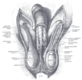
The celiac ganglia with the sympathetic plexuses of the abdominal viscera radiating from the ganglia. (Spermatic plexus labeled at right, third from the bottom.)
Details
From renal plexus
Identifiers
Latin plexus testicularis, plexus spermaticus
TA98 A14.3.03.035M
TA2 6707
FMA 6637
Anatomical terms of neuroanatomy

The spermatic plexus (or testicular plexus) is derived from the renal plexus, receiving branches from the aortic plexus. It accompanies the internal spermatic artery to the testis.

Contents

Additional images

Related Research Articles

Spermatic cord

The spermatic cord is the cord-like structure in males formed by the vas deferens and surrounding tissue that runs from the deep inguinal ring down to each testicle. Its serosal covering, the tunica vaginalis, is an extension of the peritoneum that passes through the transversalis fascia. Each testicle develops in the lower thoracic and upper lumbar region and migrates into the scrotum during its descent it carries along with it vas deferens, its vessels, nerves etc. There is one on each side.

Inguinal canal Human abdominal anatomy

The inguinal canals are the two passages in the anterior abdominal wall of humans and animals which in males convey the spermatic cords and in females the round ligament of the uterus. The inguinal canals are larger and more prominent in males. There is one inguinal canal on each side of the midline.

Genitofemoral nerve

The genitofemoral nerve refers to a nerve that is found in the abdomen. Its branches, the genital branch and femoral branch supply sensation to the upper anterior thigh, as well as the skin of the anterior scrotum in males and mons pubis in females. The femoral branch is different from the femoral nerve, which also arises from the lumbar plexus.

External iliac artery Arteries of the pelvis

The external iliac arteries are two major arteries which bifurcate off the common iliac arteries anterior to the sacroiliac joint of the pelvis.

Inferior epigastric artery Blood vessel

In human anatomy, inferior epigastric artery refers to the artery that arises from the external iliac artery. It anastomoses with the superior epigastric artery. Along its course, it is accompanied by a similarly named vein, the inferior epigastric vein. These epigastric vessels form the lateral border of Hesselbach's triangle, which outlines the area through which direct inguinal hernias protrude.

Lumbar plexus

The lumbar plexus is a web of nerves in the lumbar region of the body which forms part of the larger lumbosacral plexus. It is formed by the divisions of the first four lumbar nerves (L1-L4) and from contributions of the subcostal nerve (T12), which is the last thoracic nerve. Additionally, the ventral rami of the fourth lumbar nerve pass communicating branches, the lumbosacral trunk, to the sacral plexus. The nerves of the lumbar plexus pass in front of the hip joint and mainly support the anterior part of the thigh.

Ilioinguinal nerve

The ilioinguinal nerve is a branch of the first lumbar nerve (L1). It separates from the first lumbar nerve along with the larger iliohypogastric nerve. It emerges from the lateral border of the psoas major just inferior to the iliohypogastric, and passes obliquely across the quadratus lumborum and iliacus. The ilioinguinal nerve then perforates the transversus abdominis near the anterior part of the iliac crest, and communicates with the iliohypogastric nerve between the transversus and the internal oblique muscle.

Renal plexus

The renal plexus is formed by filaments from the celiac ganglia and plexus, aorticorenal ganglia, lower thoracic splanchnic nerves and first lumbar splanchnic nerve and aortic plexus.

Abdominal aortic plexus

The abdominal aortic plexus is formed by branches derived, on either side, from the celiac plexus and ganglia, and receives filaments from some of the lumbar ganglia.

Femoral sheath Formed by a prolongation downward, behind the inguinal ligament, of the abdominal fascia, the transversalis fascia being continued down in front of the femoral vessels and the iliac fascia behind them

The femoral sheath, also called the crural sheath, is a continuation of the abdominal fascia that is contained in the femoral triangle. It forms the femoral canal, allowing for the femoral artery and the femoral vein to travel between the abdomen and the thigh.

Testicular artery

The testicular artery is a branch of the abdominal aorta that supplies blood to the testis. It is a paired artery, with one for each of the testes.

Testicular vein

The testicular vein, the male gonadal vein, carries deoxygenated blood from its corresponding testis to the inferior vena cava or one of its tributaries. It is the male equivalent of the ovarian vein, and is the venous counterpart of the testicular artery.

Cremasteric artery

The cremasteric artery is a branch of the inferior epigastric artery which accompanies the spermatic cord, and supplies the cremaster muscle and other coverings of the cord, anastomosing with the testicular artery. In the female, the cremasteric artery is very small and accompanies the round ligament.

Medial umbilical ligament

The medial umbilical ligament is a paired structure found in human anatomy. It is on the deep surface of the anterior abdominal wall, and is covered by the medial umbilical folds. It is different from the median umbilical ligament, a structure that represents the remnant of the embryonic urachus.

Vesical nervous plexus

The vesical nervous plexus arises from the forepart of the pelvic plexus.

Muscular layer

The muscular layer is a region of muscle in many organs in the vertebrate body, adjacent to the submucosa. It is responsible for gut movement such as peristalsis. The Latin, tunica muscularis, may also be used.

External spermatic fascia

The external spermatic fascia is a thin membrane, prolonged downward around the surface of the spermatic cord and testis. It is separated from the dartos tunic by loose areolar tissue. It is occasionally referred to as 'Le Fascia de Webster' after an anatomist who once described it.

Internal spermatic fascia Thin layer around the spermatic cord

The internal spermatic fascia is a thin layer, which loosely invests the spermatic cord.

Pampiniform plexus

The pampiniform plexus is a venous plexus – a network of many small veins found in the human male spermatic cord, and the suspensory ligament of the ovary. In the male, it is formed by the union of multiple testicular veins from the back of the testis and tributaries from the epididymis.

Scrotum Anatomical male reproductive structure

The scrotum or scrotal sac is an anatomical male reproductive structure located at the base of the penis that consists of a suspended dual-chambered sac of skin and smooth muscle. It is present in most terrestrial male mammals. The scrotum contains the external spermatic fascia, testes, epididymis, and ductus deferens. It is a distention of the perineum and carries some abdominal tissues into its cavity including the testicular artery, testicular vein, and pampiniform plexus. The perineal raphe is a small, vertical, slightly raised ridge of scrotal skin under which is found the scrotal septum. It appears as a thin longitudinal line that runs front to back over the entire scrotum. In humans and some other mammals the scrotum becomes covered with pubic hair at puberty. The scrotum will usually tighten during penile erection and when exposed to cold temperatures. One testis is typically lower than the other to avoid compression in the event of an impact.

References

PD-icon.svgThis article incorporates text in the public domain from page 987 of the 20th edition of Gray's Anatomy (1918)