This article needs additional citations for verification . (November 2010) (Learn how and when to remove this template message) |
Cavernous nerve plexus | |
---|---|
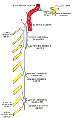
Diagram of the cervical sympathetic. (Cavernous nerve plexus labeled at top.) | |
Details | |
Identifiers | |
Latin | plexus cavernosus |
TA98 | A14.3.03.006 |
TA2 | 6650 |
FMA | 67563 |
Anatomical terms of neuroanatomy |
The cavernous nerve plexus (also called the Walther plexus [1] ) is situated below and medial to that part of the internal carotid artery which is placed by the side of the sella turcica in the cavernous sinus, and is formed chiefly by the medial division of the internal carotid nerve.
It communicates with the oculomotor, the trochlear, the ophthalmic and the abducent nerves, and with the ciliary ganglion, and distributes filaments to the wall of the internal carotid artery.
The branch of communication with the oculomotor nerve joins that nerve at its point of division; the branch to the trochlear nerve joins it as it lies on the lateral wall of the cavernous sinus; other filaments are connected with the under surface of the ophthalmic nerve; and a second filament joins the abducent nerve.
The danger triangle of the face consists of the area from the corners of the mouth to the bridge of the nose, including the nose and maxilla. Due to the special nature of the blood supply to the human nose and surrounding area, it is possible, albeit extremely unlikely, for retrograde infection from the nasal area to spread to the brain, causing cavernous sinus thrombosis, meningitis or brain abscess.
The abducens nerve is the sixth cranial nerve (CNVI), in humans, that controls the movement of the lateral rectus muscle, responsible for outward gaze. It is a somatic efferent nerve.
Articles related to anatomy include:
The oculomotor nerve is the third cranial nerve. It enters the orbit via the superior orbital fissure and innervates extrinsic eye muscles that enable most movements of the eye and that raise the eyelid. The nerve also contains fibers that innervate the intrinsic eye muscles that enable pupillary constriction and accommodation. The oculomotor nerve is derived from the basal plate of the embryonic midbrain. Cranial nerves IV and VI also participate in control of eye movement.
The internal carotid artery is located in the inner side of the neck in contrast to the external carotid artery. In human anatomy, they arise from the common carotid arteries where these bifurcate into the internal and external carotid arteries at cervical vertebral level 3 or 4; the internal carotid artery supplies the brain including eyes, while the external carotid nourishes other portions of the head, such as the face, scalp, skull, and meninges.
In anatomy, the orbit is the cavity or socket of the skull in which the eye and its appendages are situated. "Orbit" can refer to the bony socket, or it can also be used to imply the contents. In the adult human, the volume of the orbit is 30 millilitres, of which the eye occupies 6.5 ml. The orbital contents comprise the eye, the orbital and retrobulbar fascia, extraocular muscles, cranial nerves II, III, IV, V, and VI, blood vessels, fat, the lacrimal gland with its sac and duct, the eyelids, medial and lateral palpebral ligaments, check ligaments, the suspensory ligament, septum, ciliary ganglion and short ciliary nerves.
The superior orbital fissure is a foramen in the skull, although strictly it is more of a cleft, lying between the lesser and greater wings of the sphenoid bone.
The ophthalmic artery (OA) is the first branch of the internal carotid artery distal to the cavernous sinus. Branches of the OA supply all the structures in the orbit as well as some structures in the nose, face and meninges. Occlusion of the OA or its branches can produce sight-threatening conditions.
The cavernous sinus within the human head is one of the dural venous sinuses creating a cavity called the lateral sellar compartment bordered by the temporal bone of the skull and the sphenoid bone, lateral to the sella turcica.
The ophthalmic nerve (V1) is one of three divisions of the trigeminal nerve (CN V). It has three branches that provide sensory innervation to the eye, skin of the upper face and anterior scalp.
The superior ophthalmic vein begins at the inner angle of the orbit in a vein named the nasofrontal which communicates anteriorly with the angular vein; it does not pursue the same course as the ophthalmic artery and receives tributaries corresponding to the branches of that vessel.
The lesser wings of the sphenoid or orbito-sphenoids are two thin triangular plates, which arise from the upper and anterior parts of the body, and, projecting lateralward, end in sharp points [Fig. 1].
The internal carotid plexus is situated on the lateral side of the internal carotid artery, and in the plexus there occasionally exists a small gangliform swelling, the carotid ganglion, on the under surface of the artery.
The trigeminal ganglion is a sensory ganglion of the trigeminal nerve that occupies a cavity in the dura mater, covering the trigeminal impression near the apex of the petrous part of the temporal bone.
The middle cranial fossa, deeper than the anterior cranial fossa, is narrow medially and widens laterally to the sides of the skull. It is separated from the posterior fossa by the clivus and the petrous crest.
The pharyngeal branch of the vagus nerve, the principal motor nerve of the pharynx, arises from the upper part of the ganglion nodosum, and consists principally of filaments from the cranial portion of the accessory nerve.
The supratrochlear artery is one of the terminal branches of the ophthalmic artery. It branches from the ophthalmic artery near the trochlea of the superior oblique muscle in the orbit.
The following outline is provided as an overview of and topical guide to human anatomy:
The ciliary ganglion is a parasympathetic ganglion located just behind the eye in the posterior orbit. Three types of axons enter the ciliary ganglion but only the preganglionic parasympathetic axons synapse there. The entering axons are arranged into three roots of the ciliary ganglion, which join enter the posterior surface of the ganglion.
This article incorporates text in the public domain from page 978 of the 20th edition of Gray's Anatomy (1918)
This neuroscience article is a stub. You can help Wikipedia by expanding it. |