The celiac plexus or coeliac plexus, also known as the solar plexus because of its radiating nerve fibers, is a complex network of nerves located in the abdomen, near where the celiac trunk, superior mesenteric artery, and renal arteries branch from the abdominal aorta. It is behind the stomach and the omental bursa, and in front of the crura of the diaphragm, on the level of the first lumbar vertebra.

The internal anal sphincter, IAS, is a ring of smooth muscle that surrounds about 2.5–4.0 cm of the anal canal; its inferior border is in contact with, but quite separate from, the external anal sphincter.

The internal iliac artery is the main artery of the pelvis.

The inferior rectal artery is an artery that supplies blood to the lower half of the anal canal.

The abdominal aortic plexus is formed by branches derived, on either side, from the celiac plexus and ganglia, and receives filaments from some of the lumbar ganglia.

The superior hypogastric plexus is a plexus of nerves situated on the vertebral bodies anterior to the bifurcation of the abdominal aorta.

The rectal venous plexus surrounds the rectum, and communicates in front with the vesical venous plexus in the male, and the vaginal venous plexus in the female.

The superior mesenteric plexus is a continuation of the lower part of the celiac plexus, receiving a branch from the junction of the right vagus nerve with the plexus.

The superior rectal artery is an artery that descends into the pelvis to supply blood to the rectum.

The internal iliac vein begins near the upper part of the greater sciatic foramen, passes upward behind and slightly medial to the internal iliac artery and, at the brim of the pelvis, joins with the external iliac vein to form the common iliac vein.

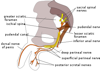
The Inferior rectal nerves usually branch from the pudendal nerve but occasionally arises directly from the sacral plexus; they cross the ischiorectal fossa along with the inferior rectal artery and veins, toward the anal canal and the lower end of the rectum, and is distributed to the Sphincter ani externus and to the integument (skin) around the anus.

The dorsal nerve of the penis is the deepest division of the pudendal nerve; it accompanies the internal pudendal artery along the ramus of the ischium; it then runs forward along the margin of the inferior ramus of the pubis, between the superior and inferior layers of the fascia of the urogenital diaphragm.

The inferior mesenteric vein begins in the rectum as the superior rectal vein, which has its origin in the hemorrhoidal plexus, and through this plexus communicates with the middle and inferior hemorrhoidal veins.

The inferior hypogastric plexus is a plexus of nerves that supplies the viscera of the pelvic cavity. The inferior hypogastric plexus gives rise to the prostatic plexus in males and the uterovaginal plexus in females.

The middle rectal veins take origin in the hemorrhoidal plexus and receive tributaries from the bladder, prostate, and seminal vesicle.

The lower part of the external hemorrhoidal plexus is drained by the inferior rectal veins into the internal pudendal vein.

The vaginal venous plexuses are placed at the sides of the vagina; they communicate with the uterine venous plexuses, vesical venous plexus, and rectal venous plexuses, and are drained by the vaginal veins, one on either side, into the hypogastric veins.

A venous plexus is a congregation of multiple veins.

The following outline is provided as an overview of and topical guide to human anatomy:
The public domain consists of all the creative works to which no exclusive intellectual property rights apply. Those rights may have expired, been forfeited, expressly waived, or may be inapplicable.

Gray's Anatomy is an English language textbook of human anatomy originally written by Henry Gray and illustrated by Henry Vandyke Carter. Earlier editions were called Anatomy: Descriptive and Surgical and Gray's Anatomy: Descriptive and Applied, but the book's name is commonly shortened to, and later editions are titled, Gray's Anatomy. The book is widely regarded as an extremely influential work on the subject, and has continued to be revised and republished from its initial publication in 1858 to the present day. The latest edition of the book, the 41st, was published in September 2015.


