The uterus or womb is the organ in the reproductive system of most female mammals, including humans, that accommodates the embryonic and fetal development of one or more embryos until birth. The uterus is a hormone-responsive sex organ that contains glands in its lining that secrete uterine milk for embryonic nourishment.

The abdominal cavity is a large body cavity in humans and many other animals that contains many organs. It is a part of the abdominopelvic cavity. It is located below the thoracic cavity, and above the pelvic cavity. Its dome-shaped roof is the thoracic diaphragm, a thin sheet of muscle under the lungs, and its floor is the pelvic inlet, opening into the pelvis.

The levator ani is a broad, thin muscle group, situated on either side of the pelvis. It is formed from three muscle components: the pubococcygeus, the iliococcygeus, and the puborectalis.

The sacrum, in human anatomy, is a large, triangular bone at the base of the spine that forms by the fusing of the sacral vertebrae (S1–S5) between ages 18 and 30.

The sigmoid colon is the part of the large intestine that is closest to the rectum and anus. It forms a loop that averages about 35–40 centimetres (14–16 in) in length. The loop is typically shaped like a Greek letter sigma (ς) or Latin letter S. This part of the colon normally lies within the pelvis, but due to its freedom of movement it is liable to be displaced into the abdominal cavity.

The pelvic floor or pelvic diaphragm is composed of muscle fibers of the levator ani, the coccygeus muscle, and associated connective tissue which span the area underneath the pelvis. The pelvic diaphragm is a muscular partition formed by the levatores ani and coccygei, with which may be included the parietal pelvic fascia on their upper and lower aspects. The pelvic floor separates the pelvic cavity above from the perineal region below. Both males and females have a pelvic floor. To accommodate the birth canal, a female's pelvic cavity is larger than a male's.

The abdomen is the part of the body between the thorax (chest) and pelvis, in humans and in other vertebrates. The abdomen is the front part of the abdominal segment of the torso. The area occupied by the abdomen is called the abdominal cavity. In arthropods it is the posterior tagma of the body; it follows the thorax or cephalothorax.

The internal iliac artery is the main artery of the pelvis. Supply Biceps femoris and quadriceps femoris

The lumbar plexus is a web of nerves in the lumbar region of the body which forms part of the larger lumbosacral plexus. It is formed by the divisions of the first four lumbar nerves (L1-L4) and from contributions of the subcostal nerve (T12), which is the last thoracic nerve. Additionally, the ventral rami of the fourth lumbar nerve pass communicating branches, the lumbosacral trunk, to the sacral plexus. The nerves of the lumbar plexus pass in front of the hip joint and mainly support the anterior part of the thigh.

The superior gluteal artery is the largest and final branch of the internal iliac artery. It is the continuation of the posterior division of that vessel. It is a short artery which runs backward between the lumbosacral trunk and the first sacral nerve. It divides into a superficial and a deep branch after passing out of the pelvis above the upper border of the piriformis muscle.

The suspensory ligament of the ovary, also infundibulopelvic ligament, is a fold of peritoneum that extends out from the ovary to the wall of the pelvis.

The pelvic cavity is a body cavity that is bounded by the bones of the pelvis. Its oblique roof is the pelvic inlet. Its lower boundary is the pelvic floor.

The pelvic brim is the edge of the pelvic inlet. It is an approximately Mickey Mouse head-shaped line passing through the prominence of the sacrum, the arcuate and pectineal lines, and the upper margin of the pubic symphysis.
The pelvic inlet or superior aperture of the pelvis is a planar surface which defines the boundary between the pelvic cavity and the abdominal cavity. It is a major target of measurements of pelvimetry.

The inferior hypogastric plexus is a network of nerves that supplies the organs of the pelvic cavity. The inferior hypogastric plexus gives rise to the prostatic plexus in males and the uterovaginal plexus in females.
Pelvic splanchnic nerves or nervi erigentes are splanchnic nerves that arise from sacral spinal nerves S2, S3, S4 to provide parasympathetic innervation to the organs of the pelvic cavity.

The pelvic fasciae are the fascia of the pelvis and can be divided into:

The following outline is provided as an overview of and topical guide to human anatomy:

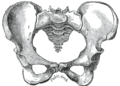
The hip bone is a large flat bone, constricted in the center and expanded above and below. In some vertebrates it is composed of three parts: the ilium, ischium, and the pubis.

The pelvis is the lower part of the trunk, between the abdomen and the thighs, together with its embedded skeleton.