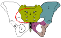
Pubis (bone)

Last updated
Pubis (bone)
Pelvic girdle illustration.svg
Pelvic girdle
Gray241.png
Male pelvis with pubis at bottom
Details
Part of Pelvis
Identifiers
Latin os pubis
MeSH D011630
TA98 A02.5.01.301
TA2 1346
FMA 16595
Anatomical terms of bone

In vertebrates, the pubis or pubic bone (Latin : os pubis) forms the lower and anterior part of each side of the hip bone. The pubis is the most forward-facing (ventral and anterior) of the three bones that make up the hip bone. The left and right pubic bones are each made up of three sections, a superior ramus, inferior ramus, and a body.

Contents

Structure

The pubic bone is made up of a body, superior ramus, and inferior ramus (Latin : branch). The left and right coxal bones join at the pubic symphysis. It is covered by a layer of fat – the mons pubis. The pubis is the lower limit of the suprapubic region. In the female, the pubis is anterior to the urethral sponge.

Body

The body of pubis has:

The body forms the wide, strong, middle and flat part of the pubic bone. The bodies of the left and right pubic bones join at the pubic symphysis. [1] The rough upper edge is the pubic crest, [2] ending laterally in the pubic tubercle. This tubercle, found roughly 3 cm from the pubic symphysis, is a distinctive feature on the lower part of the abdominal wall; important when localizing the superficial inguinal ring and the femoral canal of the inguinal canal. [3] The inner surface of the body forms part of the wall of the lesser pelvis and joints to the origin of a part of the obturator internus muscle.

Superior pubic ramus

The superior pubic ramus is the upper of the two rami. It forms the upper edge of the obturator foramen. [4] It extends from the body to the median plane where it joins with the ramus of the opposite side. It consists of an inner flattened part and a narrow outer prismoid portion.

Medial surface

Surfaces
  • The anterior surface is rough, directed downward and outward, and serves for the origin of various muscles. The adductor longus arises from the upper and medial angle, immediately below the crest; lower down, the obturator externus , the adductor brevis and the upper part of the gracilis take origin.
  • The posterior surface, convex from above downward, concave from side to side, is smooth, and forms part of the anterior wall of the pelvis. It gives origin to the levator ani and obturator internus , and attachment to the puboprostatic ligaments and to a few muscular fibers prolonged from the bladder.
Borders

The upper border presents a prominent tubercle, the pubic tubercle (pubic spine), which projects forward; the inferior crus of the subcutaneous inguinal ring (external abdominal ring), and the inguinal ligament (Poupart's ligament) are attached to it.

Passing upward and laterally from the pubic tubercle is a well-defined ridge, forming a part of the pectineal line which marks the brim of the lesser pelvis: to it are attached a portion of the inguinal falx (conjoined tendon of obliquus internus and transversus), the lacunar ligament (Gimbernat's ligament), and the reflected inguinal ligament (triangular fascia).

Medial to the pubic tubercle is the crest, which extends from this process to the medial end of the bone. It affords attachment to the inguinal falx, and to the rectus abdominis and pyramidalis .

The point of junction of the crest with the medial border of the bone is called the angle; to it, as well as to the symphysis, the superior crus of the subcutaneous inguinal ring is attached.

The medial border is articular; it is oval, and is marked by eight or nine transverse ridges, or a series of nipple-like processes arranged in rows, separated by grooves; they serve for the attachment of a thin layer of cartilage, which intervenes between it and the interpubic fibrocartilaginous lamina.

The lateral border presents a sharp margin, the obturator crest, which forms part of the circumference of the obturator foramen and affords attachment to the obturator membrane.

Lateral portion

Surfaces
  • The superior surface presents a continuation of the pectineal line, already mentioned as commencing at the pubic tubercle. In front of this line, the surface of bone is triangular in form, wider laterally than medially, and is covered by the pectineus. The surface is bounded, laterally, by a rough eminence, the iliopectineal eminence, which serves to indicate the point of junction of the ilium and pubis, and below by a prominent ridge which extends from the acetabular notch to the pubic tubercle.
  • The inferior surface forms the upper boundary of the obturator foramen, and presents, laterally, a broad and deep, oblique groove, for the passage of the obturator vessels and nerve; and medially, a sharp margin, the obturator crest, forming part of the circumference of the obturator foramen, and giving attachment to the obturator membrane.
  • The posterior surface constitutes part of the anterior boundary of the lesser pelvis. It is smooth, convex from above downward, and affords origin to some fibers of the obturator internus.

Inferior pubic ramus

The inferior pubic ramus is a part of the pelvis and is thin and flat. It passes laterally and downward from the medial end of the superior ramus; it becomes narrower as it descends and joins with the inferior ramus of the ischium below the obturator foramen.

Surfaces

Borders

  • In the female pelvis, the medial border is thick, rough, and everted, and presents two ridges, separated by an intervening space. The ridges extend downwards, and are continuous with similar ridges on the inferior ramus of the ischium;
  • The lateral border is thin and sharp, forms part of the circumference of the obturator foramen, and gives attachment to the obturator membrane.

Other animals

Mammals

Non-placental mammals possess osteological projections of the pubis known as epipubic bones. These evolved first among derived cynodonts, and evolved as a means of support for muscles flexing the thigh, facilitating the development of an erect gait. [5] However, these prevent the expansion of the torso, preventing pregnancy and forcing the animal to give birth to larval young (the modern marsupial "joeys" and monotreme "puggles"). [6]

Placentals are unique among all mammals, including other eutherians, in having lost epipubic bones and having been able to develop proper pregnancy. In some groups, remnants of these pre-pubic bones can be found as os penises. [7]

Dinosaurs

The clade Dinosauria is divided into the Saurischia and Ornithischia based on hip structure, including importantly that of the pubis. [8] An opisthopubic pelvis is a condition where the pubic bone extends back towards the tail of the animal, a trait that is also present in birds. [9] In a propubic pelvis, however, the pubic bone extends forward towards the head of the animal, as can be seen in the typical saurischian pelvic structure pictured below. The acetabulum , which can be thought of as a "hip-socket", is an opening on each side of the pelvic girdle formed where the ischium, ilium, and pubis all meet, and into which the head of the femur inserts. The orientation and position of the acetabulum is one of the main morphological traits that caused dinosaurs to walk in an upright posture with their legs directly underneath their bodies. [10] The prepubic process is a bony extension of the pubis that extends forward from the hip socket and toward the front of the animal. This adaptation is thought to have played a role in supporting the abdominal muscles.

Additional images

See also

Related Research Articles

<span class="mw-page-title-main">Human leg</span> Lower extremity or limb of the human body (foot, lower leg, thigh and hip)

The leg is the entire lower limb of the human body, including the foot, thigh or sometimes even the hip or buttock region. The major bones of the leg are the femur, tibia, and adjacent fibula. The thigh is between the hip and knee, while the calf (rear) and shin (front) are between the knee and foot.

Articles related to anatomy include:

<span class="mw-page-title-main">Pubic symphysis</span> Cartilaginous joint between the front of the left and right hip bones

The pubic symphysis is a secondary cartilaginous joint between the left and right superior rami of the pubis of the hip bones. It is in front of and below the urinary bladder. In males, the suspensory ligament of the penis attaches to the pubic symphysis. In females, the pubic symphysis is attached to the suspensory ligament of the clitoris. In most adults, it can be moved roughly 2 mm and with 1 degree rotation. This increases for women at the time of childbirth.

<span class="mw-page-title-main">Greater trochanter</span> Quadrilateral part of the thigh bone

The greater trochanter of the femur is a large, irregular, quadrilateral eminence and a part of the skeletal system.

<span class="mw-page-title-main">Piriformis muscle</span> One of six small hip muscles in the lateral rotator group

The piriformis muscle is a flat, pyramidally-shaped muscle in the gluteal region of the lower limbs. It is one of the six muscles in the lateral rotator group.

<span class="mw-page-title-main">Inguinal ligament</span> Band running from the pubic tubercle to the anterior superior iliac spine

The inguinal ligament, also known as Poupart's ligament or groin ligament, is a band running from the pubic tubercle to the anterior superior iliac spine. It forms the base of the inguinal canal through which an indirect inguinal hernia may develop.

<span class="mw-page-title-main">Obturator internus muscle</span> One of six small hip muscles in the lateral rotator group

The internal obturator muscle or obturator internus muscle originates on the medial surface of the obturator membrane, the ischium near the membrane, and the rim of the pubis.

<span class="mw-page-title-main">Hip</span> Anatomical region between the torso and the legs, holding the buttocks and genital region

In vertebrate anatomy, the hip, or coxa(pl.: coxae) in medical terminology, refers to either an anatomical region or a joint on the outer (lateral) side of the pelvis.

<span class="mw-page-title-main">External obturator muscle</span> One of six small hip muscles in the lateral rotator group

The external obturator muscle or obturator externus muscle is a flat, triangular muscle, which covers the outer surface of the anterior wall of the pelvis.

<span class="mw-page-title-main">Obturator foramen</span> Opening in the pelvis

The obturator foramen is the large, bilaterally paired opening of the bony pelvis. It is formed by the pubis and ischium. It is mostly closed by the obturator membrane except for a small opening, the obturator canal, through which the obturator nerve and vessels pass.

<span class="mw-page-title-main">Lumbar plexus</span> Web of nerves in the lower spine

The lumbar plexus is a web of nerves in the lumbar region of the body which forms part of the larger lumbosacral plexus. It is formed by the divisions of the first four lumbar nerves (L1-L4) and from contributions of the subcostal nerve (T12), which is the last thoracic nerve. Additionally, the ventral rami of the fourth lumbar nerve pass communicating branches, the lumbosacral trunk, to the sacral plexus. The nerves of the lumbar plexus pass in front of the hip joint and mainly support the anterior part of the thigh.

<span class="mw-page-title-main">Ischium</span> Lower and back region of the hip bone

The ischium forms the lower and back region of the hip bone.

<span class="mw-page-title-main">Obturator artery</span>

The obturator artery is a branch of the internal iliac artery that passes antero-inferiorly on the lateral wall of the pelvis, to the upper part of the obturator foramen, and, escaping from the pelvic cavity through the obturator canal, it divides into an anterior branch and a posterior branch.

<span class="mw-page-title-main">Upper extremity of femur</span>

The upper extremity, proximal extremity or superior epiphysis of the femur is the part of the femur closest to the pelvic bone and the trunk. It contains the following structures:

<span class="mw-page-title-main">Wing of ilium</span> Flat portion of the hip bone

The wing(ala)of ilium is the large expanded portion of the ilium, the bone which bounds the greater pelvis laterally. It presents for examination two surfaces—an external and an internal—a crest, and two borders—an anterior and a posterior.

<span class="mw-page-title-main">Pubic tubercle</span> Bone

The pubic tubercle is a prominent tubercle on the superior ramus of the pubis bone of the pelvis.

<span class="mw-page-title-main">Pelvic fascia</span>

The pelvic fasciae are the fascia of the pelvis and can be divided into:

<span class="mw-page-title-main">Outline of human anatomy</span> Overview of and topical guide to human anatomy

The following outline is provided as an overview of and topical guide to human anatomy:

<span class="mw-page-title-main">Hip bone</span> Bone of the pelvis

The hip bone is a large flat bone, constricted in the center and expanded above and below. In some vertebrates it is composed of three parts: the ilium, ischium, and the pubis.

<span class="mw-page-title-main">Pelvis</span> Lower torso of the human body

The pelvis is the lower part of the trunk, between the abdomen and the thighs, together with its embedded skeleton.

References

PD-icon.svgThis article incorporates text in the public domain from page 236 of the 20th edition of Gray's Anatomy (1918)

  1. "Definition: body of pubis from Online Medical Dictionary" . Retrieved 2008-10-18.
  2. "crista pubica" at Dorland's Medical Dictionary
  3. Bojsen-Møller, Finn (2000). Rörelseapparatens anatomi (in Swedish). Liber. p. 239. ISBN   91-47-04884-0.
  4. "Definition: superior pubic ramus from Online Medical Dictionary" . Retrieved 2008-10-18.
  5. White, T.D. (August 9, 1989). "An analysis of epipubic bone function in mammals using scaling theory". Journal of Theoretical Biology 139 (3): 343–57. doi : 10.1016/S0022-5193(89)80213-9. PMID   2615378.
  6. Michael L. Power, Jay Schulkin. The Evolution Of The Human Placenta. pp. 68–.
  7. Frederick S. Szalay (11 May 2006). Evolutionary History of the Marsupials and an Analysis of Osteological Characters. Cambridge University Press. pp. 293–. ISBN   978-0-521-02592-8.
  8. Seeley, H.G. (1888). "On the classification of the fossil animals commonly named Dinosauria." Proceedings of the Royal Society of London, 43: 165-171.
  9. Barsbold, R., (1979) Opisthopubic pelvis in the carnivorous dinosaurs. Nature. 279, 792-793
  10. Martin, A.J. (2006). Introduction to the Study of Dinosaurs. Second Edition. Oxford, Blackwell Publishing. pg. 299-300. ISBN   1-4051-3413-5.