Levator ani

Last updated
Levator ani
Gray404.png
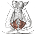
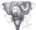
Left levator ani seen from within.
Gray1079.png
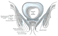
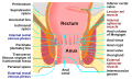
Coronal section through the male anal canal. B. Cavity of urinary bladder V.D. Vas deferens. S.V. Seminal vesicle. R. Second part of rectum. A.C. Anal canal. L.A. Levator ani. I.S. Internal anal sphincter. E.S External anal sphincter.
Details
Origin Inner surface of the side of the lesser pelvis
Insertion Inner surface of coccyx, levator ani of opposite side, and into structures that penetrate it.
Artery Inferior gluteal artery
Nerve pubococcygeus and iliococcygeus :


puborectalis :

Actions Supports the viscera in pelvic cavity
Identifiers
Latin musculus levator ani
TA98 A04.5.04.002
TA2 2403
FMA 19087
Anatomical terms of muscle
3D medical illustration presenting levator ani 3D Medical Animation Levator Ani structure.jpg
3D medical illustration presenting levator ani

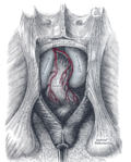
The levator ani is a broad, thin muscle group, situated on either side of the pelvis. It is formed from three muscle components: the pubococcygeus, the iliococcygeus, and the puborectalis. [3]

Contents

It is attached to the inner surface of each side of the lesser pelvis, and these unite to form the greater part of the pelvic floor. The coccygeus muscle completes the pelvic floor, which is also called the pelvic diaphragm.

It supports the viscera in the pelvic cavity, and surrounds the various structures that pass through it.

The levator ani is the main pelvic floor muscle and contracts rhythmically during female orgasm, and painfully during vaginismus. [4]

Structure

The levator ani is made up of 3 parts:

The iliococcygeus arises from the inner side of the ischium (the lower and back part of the hip bone) and from the posterior part of the tendinous arch of the obturator fascia, and is attached to the coccyx and anococcygeal body; it is usually thin, and may be absent, or be largely replaced by fibrous tissue. An accessory slip at its posterior part is sometimes named the iliosacralis.

The pubococcygeus muscle has medial fibres forming the pubovaginalis in the female, and the puboprostaticus in the male.

Origin and insertion

The levator ani arises, in front, from the posterior surface of the superior pubic ramus lateral to the symphysis; behind, from the inner surface of the spine of the ischium; and between these two points, from the obturator fascia.

Posteriorly, this fascial origin corresponds, more or less closely, with the tendinous arch of the pelvic fascia, but in front, the muscle arises from the fascia at a varying distance above the arch, in some cases reaching nearly as high as the canal for the obturator vessels and nerve.

The fibers pass downward and backward to the middle line of the floor of the pelvis; the most posterior are inserted into the side of the last two segments of the coccyx; those placed more anteriorly unite with the muscle of the opposite side, in a median fibrous ridge called the anococcygeal body or raphe, which extends between the coccyx and the margin of the anus.

The middle fibers are inserted into the side of the rectum, blending with the fibers of the sphincter muscles; lastly in the male, the anterior fibers descend upon the side of the prostate to unite beneath it with the muscle of the opposite side, joining with the fibers of the external anal sphincter and transverse perineal muscles, at the central tendinous point of the perineum.

The anterior portion is occasionally separated from the rest of the muscle by connective tissue.

From this circumstance, as well as from its peculiar relation with the prostate, which it supports as in a sling, it has been described as a distinct muscle, under the name of levator prostatæ.

In the female the anterior fibers of the levator ani descend upon the side of the vagina.

Innervation

The levator ani muscles are mostly innervated by the pudendal nerve, perineal nerve and acting together. [5]

Variation

In addition, sacral spinal nerves (S3, S4) innervate the muscles directly as well (in ~70% [5] of people). Sometimes (in ~40% [5] of people) the inferior rectal nerve innervates the levator ani muscles independently of the pudendal nerve.

Pubococcygeus muscle

Pubococcygeus muscle
Details
Origin back of the pubis and from the anterior part of the obturator fascia
Insertion coccyx and sacrum
Nerve S3, S4
Actions controls urine flow and contracts during orgasm
Identifiers
Latin musculus pubococcygeus
TA98 A04.5.04.002
TA2 2403
FMA 19087
Anatomical terms of muscle

The pubococcygeus muscle or PC muscle is a hammock-like muscle, found in both sexes, that stretches from the pubic bone to the coccyx (tail bone) forming the floor of the pelvic cavity and supporting the pelvic organs.

Structure

The pubococcygeus arises from the back of the pubis and from the anterior part of the obturator fascia, and is directed backward almost horizontally along the side of the anal canal toward the coccyx and sacrum, to which it finds attachment.

Between the termination of the vertebral column and the anus, the two pubococcygeus muscles come together and form a thick, fibromuscular layer lying on the raphe (ridge) or (anococcygeal body) formed by the iliococcygei.

The greater part of this muscle is inserted into the coccyx and into the last one or two pieces of the sacrum.

Variation

This insertion into the vertebral column is, however, not accepted by all observers.

Function

The pubococcygeus muscle controls urine flow and contracts during orgasm as well as assisting in male ejaculation. [6] It also aids in childbirth as well as core stability. [7]

A strong pubococcygeus muscle has also been linked to a reduction in urinary incontinence and proper positioning of the baby's head during childbirth.

Kegel exercises

The Kegel exercises are a series of voluntary contractions of all the perineal muscles. Such movement is done in an effort to strengthen all the striated muscles in the perineum's area. They are often referred to simply as "kegels", named after their founder, Dr. Arnold Kegel. [8] These exercises also serve to contract, among others, the ischiocavernosus, bulbospongiosus, and cremaster muscle in men, as voluntary contraction of the pubococcygeus muscle also engages the cremasteric reflex, which lifts the testicles up, although this does not occur in all men. Kegel exercises have been prescribed to ameliorate erectile dysfunction due to venous leakage and to help men control premature ejaculation [9] and to treat urinary incontinence in both sexes. [10] [11]

Puborectalis muscle

Puborectalis muscle
Gray1077.png
The posterior aspect of the rectum exposed by removing the lower part of the sacrum and the coccyx. (Puborectalis not labeled but levator ani labeled at bottom right and external anal sphincter labeled at bottom center.)
Details
Origin lower part of the pubic symphysis, superior fascia of the urogenital diaphragm
Nerve S3, S4. levator ani nerve [12]
Actions inhibit defecation
Identifiers
Latin musculus puborectalis
TA98 A04.5.04.002
TA2 2403
FMA 19087
Anatomical terms of muscle

The fibers that form a sling looping around the rectum are named the puborectalis. They arise from the lower part of the pubic symphysis, and from the superior fascia of the urogenital diaphragm. The origin of the puborectalis fibers is at the posterior surface of the pubis while their insertion is at the midline sling posterior to the rectum. The muscle band is innervated by perineal branches of the S3 and S4 nerve roots.

They meet with the corresponding fibers of the opposite side around the lower part of the rectum, and form for it a strong sling. Relaxation increases the angle between rectum and anus, allowing defecation in conjunction with relaxation of the internal and external anal sphincters. Levator ani relaxation and rectal emptying is facilitated by anorectal straightening during squatting. [13]

Function

The levator ani muscles are responsible for "wagging" the tail in tailed quadrupeds. These muscles are not as strong in the human, as tail-wagging is more demanding than the support function that the muscles serve in humans. [14]

Clinical significance

Levator ani syndrome

Levator ani syndrome is episodic rectal pain caused by spasm of the levator ani muscle. [15] [16] [17]

Levator ani avulsion

[18]

Additional images

See also

Related Research Articles

<span class="mw-page-title-main">Perineum</span> Region of the body including the perineal body and surrounding structures

The perineum in humans is the space between the anus and scrotum in the male or between the anus and vulva in the female. The perineum is the region of the body between the pubic symphysis and the coccyx, including the perineal body and surrounding structures. The perineal raphe is visible and pronounced to varying degrees. The perineum is an erogenous zone. This area is also known as the taint or gooch in American slang.

<span class="mw-page-title-main">Pudendal nerve</span> Main nerve of the perineum

The pudendal nerve is the main nerve of the perineum. It is a mixed nerve and also conveys sympathetic autonomic fibers. It carries sensation from the external genitalia of both sexes and the skin around the anus and perineum, as well as the motor supply to various pelvic muscles, including the male or female external urethral sphincter and the external anal sphincter.

<span class="mw-page-title-main">Coccyx</span> Bone of the pelvis

The coccyx, commonly referred to as the tailbone, is the final segment of the vertebral column in all apes, and analogous structures in certain other mammals such as horses. In tailless primates since Nacholapithecus, the coccyx is the remnant of a vestigial tail. In animals with bony tails, it is known as tailhead or dock, in bird anatomy as tailfan. It comprises three to five separate or fused coccygeal vertebrae below the sacrum, attached to the sacrum by a fibrocartilaginous joint, the sacrococcygeal symphysis, which permits limited movement between the sacrum and the coccyx.

<span class="mw-page-title-main">Kegel exercise</span> Pelvic floor exercise

Kegel exercise, also known as pelvic floor exercise, involves repeatedly contracting and relaxing the muscles that form part of the pelvic floor, now sometimes colloquially referred to as the "Kegel muscles". The exercise can be performed many times a day, for several minutes at a time but takes one to three months to begin to have an effect.

<span class="mw-page-title-main">Bulbospongiosus muscle</span> Superficial muscle of the perineum

The bulbospongiosus muscle is one of the superficial muscles of the perineum. It has a slightly different origin, insertion and function in males and females. In males, it covers the bulb of the penis. In females, it covers the vestibular bulb.

<span class="mw-page-title-main">Pelvic floor</span> Anatomical structure

The pelvic floor or pelvic diaphragm is an anatomical location in the human body, which has an important role in urinary and anal continence, sexual function and support of the pelvic organs. The pelvic floor includes muscles, both skeletal and smooth, ligaments and fascia. and separates between the pelvic cavity from above, and the perineum from below. It is formed by the levator ani muscle and coccygeus muscle, and associated connective tissue.

<span class="mw-page-title-main">Pectineus muscle</span> Adductor of the thigh

The pectineus muscle is a flat, quadrangular muscle, situated at the anterior (front) part of the upper and medial (inner) aspect of the thigh. The pectineus muscle is the most anterior adductor of the hip. The muscle's primary action is hip flexion; it also produces adduction and internal rotation of the hip.

<span class="mw-page-title-main">Coccygeus muscle</span> Muscle of the lower back arising by its apex from the spine of the ischium

The coccygeus muscle or ischiococcygeus is a muscle of the pelvic floor, located posterior to levator ani and anterior to the sacrospinous ligament.

<span class="mw-page-title-main">Obturator internus muscle</span> One of six small hip muscles in the lateral rotator group

The internal obturator muscle or obturator internus muscle originates on the medial surface of the obturator membrane, the ischium near the membrane, and the rim of the pubis.

<span class="mw-page-title-main">External anal sphincter</span> Flat plane of skeletal muscle fibers

The external anal sphincter is an oval tube of skeletal muscle fibers. Distally, it is adherent to the skin surrounding the margin of the anus. It exhibits a resting state of tonical contraction.

<span class="mw-page-title-main">Pubis (bone)</span> Most forward-facing of the three main regions making up the os coxa

In vertebrates, the pubis or pubic bone forms the lower and anterior part of each side of the hip bone. The pubis is the most forward-facing of the three bones that make up the hip bone. The left and right pubic bones are each made up of three sections, a superior ramus, inferior ramus, and a body.

<span class="mw-page-title-main">Sacrotuberous ligament</span>

The sacrotuberous ligament is situated at the lower and back part of the pelvis. It is flat, and triangular in form; narrower in the middle than at the ends.

<span class="mw-page-title-main">Pelvic cavity</span> Body cavity bounded by the pelvic bones

The pelvic cavity is a body cavity that is bounded by the bones of the pelvis. Its oblique roof is the pelvic inlet. Its lower boundary is the pelvic floor.

<span class="mw-page-title-main">Obturator fascia</span>

The obturator fascia, or fascia of the internal obturator muscle, covers the pelvic surface of that muscle and is attached around the margin of its origin.

<span class="mw-page-title-main">Deep perineal pouch</span> Anatomic space enclosed partly by the perineum

The deep perineal pouch is the anatomic space enclosed in part by the perineum and located superior to the perineal membrane.

<span class="mw-page-title-main">Urogenital triangle</span> Anterior part of the perineum

The urogenital triangle is the anterior part of the perineum. In female mammals, it contains the vagina and associated parts of the internal genitalia.

<span class="mw-page-title-main">Anal triangle</span> Posterior part of the perineum

The anal triangle is the posterior part of the perineum. It contains the anal canal.

<span class="mw-page-title-main">Pelvic fascia</span>

The pelvic fasciae are the fascia of the pelvis and can be divided into:

<span class="mw-page-title-main">Defecography</span> Visualisation of the mechanics of a patients defecation

Defecography is a type of medical radiological imaging in which the mechanics of a patient's defecation are visualized in real time using a fluoroscope. The anatomy and function of the anorectum and pelvic floor can be dynamically studied at various stages during defecation.

<span class="mw-page-title-main">Vaginal support structures</span> Structures that maintain the position of the vagina within the pelvic cavity

The vaginal support structures are those muscles, bones, ligaments, tendons, membranes and fascia, of the pelvic floor that maintain the position of the vagina within the pelvic cavity and allow the normal functioning of the vagina and other reproductive structures in the female. Defects or injuries to these support structures in the pelvic floor leads to pelvic organ prolapse. Anatomical and congenital variations of vaginal support structures can predispose a woman to further dysfunction and prolapse later in life. The urethra is part of the anterior wall of the vagina and damage to the support structures there can lead to incontinence and urinary retention.

References

PD-icon.svgThis article incorporates text in the public domain from page 422 of the 20th edition of Gray's Anatomy (1918)

  1. Essential Clinical Anatomy. K.L. Moore & A.M. Agur. Lippincott, 2nd ed. 2002. Page 217
  2. Wallner C, Maas C, Dabhoiwala N, Lamers W, Deruiter M (2006). "Evidence for the innervation of the puborectalis muscle by the levator ani nerve". Neurogastroenterol Motil. 18 (12): 1121–1122. doi:10.1111/j.1365-2982.2006.00846.x. PMID   17109696. S2CID   29088779.
  3. Drake, Richard; Vogl, A. Wayne; Mitchell, Adam (2015). Gray's Anatomy for Students (Third ed.). Elsevier.
  4. Christopher R. Chapple (2006). Multidisciplinary Management of Female Pelvic Floor Disorders. Churchill Livingstone. pp. 4–. ISBN   978-0-443-07272-7.
  5. 1 2 3 Grigorescu BA, Lazarou G, Olson TR, et al. (2008). "Innervation of the levator ani muscles: description of the nerve branches to the pubococcygeus, iliococcygeus, and puborectalis muscles". Int Urogynecol J Pelvic Floor Dysfunct. 19 (1): 107–116. doi:10.1007/s00192-007-0395-8. PMID   17565421. S2CID   21118078.
  6. Marieb, Elaine (2013). Anatomy & physiology : books a la carte edition. Benjamin-Cummings. p. 895. ISBN   9780321887603.
  7. "Core stabilization, Core Coordination, article by Aline Newton, M.A., certified Advanced Rolfer, the biomechanics of core stabilization, with core as a center of movement instead of a center of holding". Alinenewton.com. Retrieved 2012-02-20.
  8. Kegel exercises Gannet Health Services. Cornwell University website
  9. How To do Kegel Exercises (for men) Silverberg, Corey. About.com
  10. Dumoulin, Chantale; Hay-Smith, E. Jean C.; Mac Habée-Séguin, Gabrielle (2014-05-14). "Pelvic floor muscle training versus no treatment, or inactive control treatments, for urinary incontinence in women". The Cochrane Database of Systematic Reviews (5): CD005654. doi:10.1002/14651858.CD005654.pub3. ISSN   1469-493X. PMID   24823491.
  11. Anderson, Coral A.; Omar, Muhammad Imran; Campbell, Susan E.; Hunter, Kathleen F.; Cody, June D.; Glazener, Cathryn M. A. (2015-01-20). "Conservative management for postprostatectomy urinary incontinence". The Cochrane Database of Systematic Reviews. 1 (1): CD001843. doi:10.1002/14651858.CD001843.pub5. hdl: 2164/6141 . ISSN   1469-493X. PMC   7025637 . PMID   25602133.
  12. Wallner C, Maas C, Dabhoiwala N, Lamers W, Deruiter M (2006). "Evidence for the innervation of the puborectalis muscle by the levator ani nerve". Neurogastroenterol Motil. 18 (12): 1121–2. doi:10.1111/j.1365-2982.2006.00846.x. PMID   17109696. S2CID   29088779.
  13. Modi, Rohan M.; Hinton, Alice; Pinkhas, Daniel; Groce, Royce; Meyer, Marty M.; Balasubramanian, Gokulakrishnan; Levine, Edward; Stanich, Peter P. (March 2019). "Implementation of a Defecation Posture Modification Device". Journal of Clinical Gastroenterology. 53 (3): 216–219. doi:10.1097/MCG.0000000000001143. ISSN   0192-0790. PMC   6382038 . PMID   30346317.
  14. Sloan, Ethel (2001, p. 53). Biology of Women, Wisconsin: CENGAGE Delmar Learning. ISBN   0-7668-1142-5 (excerpt available here Archived 2006-05-21 at the Wayback Machine )
  15. Levator Syndrome, by Parswa Ansari, MD 7/2014, Merck Manuals
  16. Giulio Aniello Santoro; Andrzej Paweł Wieczorek; Clive I. Bartram (27 October 2010). Pelvic Floor Disorders: Imaging and Multidisciplinary Approach to Management. Springer. p. 601. ISBN   978-88-470-1542-5.
  17. Bharucha AE, Trabuco E (September 2008). "Functional and chronic anorectal and pelvic pain disorders". Gastroenterology Clinics of North America. 37 (3): 685–96, ix. doi:10.1016/j.gtc.2008.06.002. PMC   2676775 . PMID   18794003.
  18. Rusavy, Z; Paymova, L; Kozerovsky, M; Veverkova, A; Kalis, V; Kamel, RA; Ismail, KM (2021). "Levator ani avulsion: a Systematic evidence review (LASER)". BJOG: An International Journal of Obstetrics & Gynaecology. Wiley. 129 (4): 517–528. doi:10.1111/1471-0528.16837. ISSN   1470-0328. PMID   34245656.