Conjoint tendon

Last updated
Conjoint tendon
Gray398.png
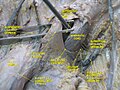
The interfoveolar ligament, seen from in front. (Inguinal aponeurotic falx labeled at lower left.)
Details
Identifiers
Latin falx inguinalis, tendo conjunctivus
TA98 A04.5.01.020
TA2 2376
FMA 20275
Anatomical terminology

The conjoint tendon (previously known as the inguinal aponeurotic falx) is a sheath of connective tissue formed from the lower part of the common aponeurosis of the abdominal internal oblique muscle and the transversus abdominis muscle, joining the muscle to the pelvis. It forms the medial part of the posterior wall of the inguinal canal.

Contents

Structure

The conjoint tendon is formed from the lower part of the common aponeurosis of the abdominal internal oblique muscle and the transversus abdominis muscle. [1] It inserts into the pubic crest and the pectineal line immediately behind the superficial inguinal ring. [1] It is usually conjoint with the tendon of the internal oblique muscle, but they may be separate as well. It forms the medial part of the posterior wall of the inguinal canal. [1]

Clinical significance

The conjoint tendon serves to protect what would otherwise be a weak point in the abdominal wall. [1] A weakening of the conjoint tendon can precipitate a direct inguinal hernia. [1] [2]

A direct inguinal hernia will protrude through Hesselbach's triangle, whose borders are the rectus abdominis (medially), inferior epigastric artery and inferior epigastric vein (superolaterally), and the inguinal ligament (inferiorly). The hernia lies medial to the inferior epigastric artery. [3] This is in contrast to an indirect inguinal hernia, which will protrude laterally to the inferior epigastric artery and is most commonly due to an embryological defect in the closure of the deep inguinal ring.

History

The conjoint tendon is also known as the inguinal aponeurotic falx, and Henle's ligament. [4]

Additional images

See also

Related Research Articles

<span class="mw-page-title-main">Inguinal canal</span> Human abdominal anatomy

The inguinal canal is a passage in the anterior abdominal wall on each side of the body, which in males, convey the spermatic cords and in females, the round ligament of the uterus. The inguinal canals are larger and more prominent in males.

<span class="mw-page-title-main">Aponeurosis</span> Tissue which connects muscles to other organs

An aponeurosis is a flattened tendon by which muscle attaches to bone or fascia. Aponeuroses exhibit an ordered arrangement of collagen fibres, thus attaining high tensile strength in a particular direction while being vulnerable to tensional or shear forces in other directions. They have a shiny, whitish-silvery color, are histologically similar to tendons, and are very sparingly supplied with blood vessels and nerves. When dissected, aponeuroses are papery and peel off by sections. The primary regions with thick aponeuroses are in the ventral abdominal region, the dorsal lumbar region, the ventriculus in birds, and the palmar (palms) and plantar (soles) regions.

<span class="mw-page-title-main">Rectus abdominis muscle</span> Paired straight muscle

The rectus abdominis muscle, also known as the "abdominal muscle" or simply the "abs", is a pair of segmented skeletal muscle on the ventral aspect of a person's abdomen. The paired muscle is separated at the midline by a band of dense connective tissue called the linea alba, and the connective tissue defining each lateral margin of the rectus abdominus is the linea semilunaris. The muscle extends from the pubic symphysis, pubic crest and pubic tubercle inferiorly, to the xiphoid process and costal cartilages of the 5th–7th ribs superiorly.

<span class="mw-page-title-main">Inguinal ligament</span> Band running from the pubic tubercle to the anterior superior iliac spine

The inguinal ligament, also known as Poupart's ligament or groin ligament, is a band running from the pubic tubercle to the anterior superior iliac spine. It forms the base of the inguinal canal through which an indirect inguinal hernia may develop.

<span class="mw-page-title-main">Transverse abdominal muscle</span> Muscle of the abdominal area

The transverse abdominal muscle (TVA), also known as the transverse abdominis, transversalis muscle and transversus abdominis muscle, is a muscle layer of the anterior and lateral abdominal wall, deep to the internal oblique muscle. It is thought by most fitness instructors to be a significant component of the core.

<span class="mw-page-title-main">External iliac artery</span> Arteries of the pelvis

The external iliac arteries are two major arteries which bifurcate off the common iliac arteries anterior to the sacroiliac joint of the pelvis.

<span class="mw-page-title-main">Abdominal external oblique muscle</span> Skeletal muscle in the abdomen

The abdominal external oblique muscle is the largest and outermost of the three flat abdominal muscles of the lateral anterior abdomen.

<span class="mw-page-title-main">Inferior epigastric artery</span> Blood vessel

In human anatomy, the inferior epigastric artery is an artery that arises from the external iliac artery. It is accompanied by the inferior epigastric vein; inferiorly, these two inferior epigastric vessels together travel within the lateral umbilical fold The inferior epigastric artery then traverses the arcuate line of rectus sheath to enter the rectus sheath, then anastomoses with the superior epigastric artery within the rectus sheath.

<span class="mw-page-title-main">Lumbar plexus</span> Web of nerves in the lower spine

The lumbar plexus is a web of nerves in the lumbar region of the body which forms part of the larger lumbosacral plexus. It is formed by the divisions of the first four lumbar nerves (L1-L4) and from contributions of the subcostal nerve (T12), which is the last thoracic nerve. Additionally, the ventral rami of the fourth lumbar nerve pass communicating branches, the lumbosacral trunk, to the sacral plexus. The nerves of the lumbar plexus pass in front of the hip joint and mainly support the anterior part of the thigh.

<span class="mw-page-title-main">Ilioinguinal nerve</span> Branch of the first lumbar nerve

The ilioinguinal nerve is a branch of the first lumbar nerve (L1). It separates from the first lumbar nerve along with the larger iliohypogastric nerve. It emerges from the lateral border of the psoas major just inferior to the iliohypogastric, and passes obliquely across the quadratus lumborum and iliacus. The ilioinguinal nerve then perforates the transversus abdominis near the anterior part of the iliac crest, and communicates with the iliohypogastric nerve between the transversus and the internal oblique muscle.

<span class="mw-page-title-main">Pubis (bone)</span> Most forward-facing of the three main regions making up the os coxa

In vertebrates, the pubis or pubic bone forms the lower and anterior part of each side of the hip bone. The pubis is the most forward-facing of the three bones that make up the hip bone. The left and right pubic bones are each made up of three sections, a superior ramus, inferior ramus, and a body.

In human anatomy, the inguinal region refers to either the groin or the lower lateral regions of the abdomen. It may also refer to:

<span class="mw-page-title-main">Pubic tubercle</span> Bone

The pubic tubercle is a prominent tubercle on the superior ramus of the pubis bone of the pelvis.

The subcostal arteries, so named because they lie below the last ribs, constitute the lowest pair of branches derived from the thoracic aorta, and are in series with the intercostal arteries.

<span class="mw-page-title-main">Lateral umbilical fold</span>

The lateral umbilical fold is an elevation of the peritoneum lining the inner/posterior surface of the lower anterior abdominal wall formed by the underlying inferior epigastric artery and inferior epigastric vein which the peritoneum covers. Superiorly, the lateral umbilical fold ends where the vessels reach and enter the rectus sheath at the arcuate line of rectus sheath; in spite of the name, the lateral umbilical folds do not extend as far superiorly as the umbilicus. Inferiorly, it extends to just medial to the deep inguinal ring.

<span class="mw-page-title-main">Arcuate line of rectus sheath</span> Line of demarcation in the human abdomen

The arcuate line of rectus sheath is a line of demarcation corresponding to the free inferior margin of the posterior layer of the rectus sheath inferior to which only the anterior layer of the rectus sheath is present and the rectus abdominis muscle is therefore in direct contact with the transversalis fascia. The arcuate line is concave inferior-wards.

<span class="mw-page-title-main">Medial umbilical ligament</span>

The medial umbilical ligament, cord of umbilical artery, or obliterated umbilical artery is a paired structure found in human anatomy. It is on the deep surface of the anterior abdominal wall, and is covered by the medial umbilical folds. It is different from the median umbilical ligament, a structure that represents the remnant of the embryonic urachus.

<span class="mw-page-title-main">Deep circumflex iliac artery</span> Artery in the pelvis

The deep circumflex iliac artery is an artery in the pelvis that travels along the iliac crest of the pelvic bone.

<span class="mw-page-title-main">Rectus sheath</span> Laminas around abdominal muscles

The rectus sheath is a tough fibrous compartment formed by the aponeuroses of the transverse abdominal muscle, and the internal and external oblique muscles. It contains the rectus abdominis and pyramidalis muscles, as well as vessels and nerves.

<span class="mw-page-title-main">Outline of human anatomy</span> Overview of and topical guide to human anatomy

The following outline is provided as an overview of and topical guide to human anatomy:

References

  1. 1 2 3 4 5 Jiang, Jay; Koay, Jennifer (2021), "Anatomy, Abdomen and Pelvis, Conjoint Tendon (Inguinal Aponeurotic Falx)", StatPearls, Treasure Island (FL): StatPearls Publishing, PMID   31747179 , retrieved 2021-04-12
  2. Relevant Anatomy Archived 2012-12-30 at the Wayback Machine at University of Connecticut Health Center. Retrieved Jan 2013
  3. Clinical Anatomy by Ernest W. April. 3rd Edition. Published by Lippincott Williams & Wilkins, 1997. Pages 326-327.
  4. Jiang, Jay; Koay, Jennifer (2021), "Anatomy, Abdomen and Pelvis, Conjoint Tendon (Inguinal Aponeurotic Falx)", StatPearls, Treasure Island (FL): StatPearls Publishing, PMID   31747179 , retrieved 2021-04-12