Inferior epigastric artery

Last updated

Inferior epigastric artery
Gray547.png
Right inferior epigastric artery - view from inside of abdomen. (Inferior epigastric vessels labeled at upper left.)
Gray401.png
The abdominal inguinal ring. (Inf. epigastric artery labeled at center left.)
Details
Source External iliac artery
Branches Cremasteric artery, pubic branch of inferior epigastric artery, artery of round ligament of uterus
Vein Inferior epigastric vein
Identifiers
Latin arteria epigastrica inferior
TA98 A12.2.16.003
TA2 4358
FMA 20686
Anatomical terminology

In human anatomy, the inferior epigastric artery is an artery that arises from the external iliac artery. [1] :225 It is accompanied by the inferior epigastric vein; inferiorly, these two inferior epigastric vessels together travel within the lateral umbilical fold (which represents the lateral border of Hesselbach's triangle, the area through which direct inguinal hernias protrude. [2] ) The inferior epigastric artery then traverses the arcuate line of rectus sheath to enter the rectus sheath, [1] :234 then anastomoses with the superior epigastric artery within the rectus sheath. [1] :225

Contents

Structure

Origin

The inferior epigastric artery arises from the external iliac artery, immediately superior to the inguinal ligament. [3]

Course and relations

It curves forward in the subperitoneal tissue, and then ascends obliquely along the medial margin of the abdominal inguinal ring; continuing its course upward, it pierces the transversalis fascia, and, passing in front of the linea semicircularis, ascends between the rectus abdominis muscle and the posterior lamella of its sheath.

It finally divides into numerous branches, which anastomose, above the umbilicus, with the superior epigastric branch of the internal thoracic artery and with the lower intercostal arteries.

As the inferior epigastric artery passes obliquely upward from its origin it lies along the lower and medial margins of the abdominal inguinal ring, and behind the commencement of the spermatic cord.

The vas deferens, as it leaves the spermatic cord in the male, and the round ligament of the uterus in the female, winds around the lateral and posterior aspects of the artery.

Anastomoses

It anastomoses with the superior epigastric artery. [3]

Clinical significance

Hernia

The inferior epigastric artery may lie close to an inguinal hernia, so acts as a useful landmark. [4]

Surgery

The inferior epigastric artery may be damaged during laparoscopic surgery. [4] It may also be damaged when manually finding the peritoneum beneath the rectus abdominis muscle. [4]

Additional images

See also

Related Research Articles

<span class="mw-page-title-main">Inguinal canal</span> Human abdominal anatomy

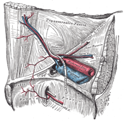
The inguinal canal is a passage in the anterior abdominal wall on each side of the body, which in males, convey the spermatic cords and in females, the round ligament of the uterus. The inguinal canals are larger and more prominent in males.

<span class="mw-page-title-main">Rectus abdominis muscle</span> Paired straight muscle

The rectus abdominis muscle, also known as the "abdominal muscle" or simply the "abs", is a pair of segmented skeletal muscle on the ventral aspect of a person's abdomen. The paired muscle is separated at the midline by a band of dense connective tissue called the linea alba, and the connective tissue defining each lateral margin of the rectus abdominus is the linea semilunaris. The muscle extends from the pubic symphysis, pubic crest and pubic tubercle inferiorly, to the xiphoid process and costal cartilages of the 5th–7th ribs superiorly.

<span class="mw-page-title-main">Inguinal ligament</span> Band running from the pubic tubercle to the anterior superior iliac spine

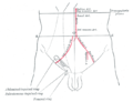
The inguinal ligament, also known as Poupart's ligament or groin ligament, is a band running from the pubic tubercle to the anterior superior iliac spine. It forms the base of the inguinal canal through which an indirect inguinal hernia may develop.

<span class="mw-page-title-main">Transverse abdominal muscle</span> Muscle of the abdominal area

The transverse abdominal muscle (TVA), also known as the transverse abdominis, transversalis muscle and transversus abdominis muscle, is a muscle layer of the anterior and lateral abdominal wall, deep to the internal oblique muscle. It is thought by most fitness instructors to be a significant component of the core.

<span class="mw-page-title-main">External iliac artery</span> Arteries of the pelvis

The external iliac arteries are two major arteries which bifurcate off the common iliac arteries anterior to the sacroiliac joint of the pelvis.

<span class="mw-page-title-main">Internal iliac artery</span> Main artery of the pelvis

The internal iliac artery is the main artery of the pelvis.

<span class="mw-page-title-main">Inguinal triangle</span> Region of the abdominal wall in humans

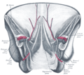
In human anatomy, the inguinal triangle is a region of the abdominal wall. It is also known by the eponym Hesselbach's triangle, after Franz Kaspar Hesselbach.

<span class="mw-page-title-main">Inferior epigastric vein</span> Large blood vessel

In human anatomy, inferior epigastric vein are 1-2 veins accompanying the inferior epigastric artery. They drain into the external iliac vein just proximal to the inguinal ligament.

<span class="mw-page-title-main">Superior epigastric artery</span> Blood vessel

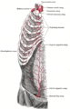
In human anatomy, the superior epigastric artery is a terminal branch of the internal thoracic artery that provides arterial supply to the abdominal wall, and upper rectus abdominis muscle. It enters the rectus sheath to descend upon the inner surface of the rectus abdominis muscle. It ends by anastomosing with the inferior epigastric artery.

<span class="mw-page-title-main">Superior epigastric vein</span> Blood vessel

In human anatomy, the superior epigastric veins are two or more venae comitantes which accompany either superior epigastric artery before emptying into the internal thoracic vein. They participate in the drainage of the superior surface of the diaphragm.

<span class="mw-page-title-main">Conjoint tendon</span> Medial part of the posterior wall of the inguinal canal

The conjoint tendon is a sheath of connective tissue formed from the lower part of the common aponeurosis of the abdominal internal oblique muscle and the transversus abdominis muscle, joining the muscle to the pelvis. It forms the medial part of the posterior wall of the inguinal canal.

<span class="mw-page-title-main">Lumbar arteries</span> Arteries within the lower back

The lumbar arteries are arteries located in the lower back or lumbar region. The lumbar arteries are in parallel with the intercostals.

In human anatomy, the inguinal region refers to either the groin or the lower lateral regions of the abdomen. It may also refer to:

<span class="mw-page-title-main">Transversalis fascia</span> Aponeurosis between the transverse abdominal muscle and the extraperitoneal fat

The transversalis fascia is the fascial lining of the anterolateral abdominal wall situated between the inner surface of the transverse abdominal muscle, and the preperitoneal fascia. It is directly continuous with the iliac fascia, the internal spermatic fascia, and pelvic fascia.

<span class="mw-page-title-main">Lateral umbilical fold</span>

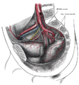
The lateral umbilical fold is an elevation of the peritoneum lining the inner/posterior surface of the lower anterior abdominal wall formed by the underlying inferior epigastric artery and inferior epigastric vein which the peritoneum covers. Superiorly, the lateral umbilical fold ends where the vessels reach and enter the rectus sheath at the arcuate line of rectus sheath; in spite of the name, the lateral umbilical folds do not extend as far superiorly as the umbilicus. Inferiorly, it extends to just medial to the deep inguinal ring.

<span class="mw-page-title-main">Testicular artery</span> Branch of the abdominal aorta that supplies blood to the testicle

The testicular artery is a branch of the abdominal aorta that supplies blood to the testicle. It is a paired artery, with one for each of the testicles.

<span class="mw-page-title-main">Arcuate line of rectus sheath</span> Line of demarcation in the human abdomen

The arcuate line of rectus sheath is a line of demarcation corresponding to the free inferior margin of the posterior layer of the rectus sheath inferior to which only the anterior layer of the rectus sheath is present and the rectus abdominis muscle is therefore in direct contact with the transversalis fascia. The arcuate line is concave inferior-wards.

<span class="mw-page-title-main">Medial umbilical ligament</span>

The medial umbilical ligament, cord of umbilical artery, or obliterated umbilical artery is a paired structure found in human anatomy. It is on the deep surface of the anterior abdominal wall, and is covered by the medial umbilical folds. It is different from the median umbilical ligament, a structure that represents the remnant of the embryonic urachus.

<span class="mw-page-title-main">Deep circumflex iliac artery</span> Artery in the pelvis

The deep circumflex iliac artery is an artery in the pelvis that travels along the iliac crest of the pelvic bone.

<span class="mw-page-title-main">Rectus sheath</span> Laminas around abdominal muscles

The rectus sheath is a tough fibrous compartment formed by the aponeuroses of the transverse abdominal muscle, and the internal and external oblique muscles. It contains the rectus abdominis and pyramidalis muscles, as well as vessels and nerves.

References

PD-icon.svgThis article incorporates text in the public domain from page 623 of the 20th edition of Gray's Anatomy (1918)

  1. 1 2 3 Sinnatamby C (2011). Last's Anatomy (12th ed.). ISBN   978-0-7295-3752-0.
  2. Wu WT, Chang KV, Lin CP, Yeh CC, Özçakar L (July 2022). "Ultrasound imaging for inguinal hernia: a pictorial review". Ultrasonography. 41 (3): 610–623. doi:10.14366/usg.21192. PMC   9262670 . PMID   35569836.
  3. 1 2 Castro Ferreira M, Henrique Ishida L, Munhoz A (January 2009). "CHAPTER 19 - Rectus flap". In Wei FC, Mardini S (eds.). Flaps and Reconstructive Surgery. Edinburgh: W.B. Saunders. pp. 207–223. doi:10.1016/B978-0-7216-0519-7.00019-8. ISBN   978-0-7216-0519-7.
  4. 1 2 3 Paterson-Brown S (January 2010). "Chapter Five - Applied anatomy". In Bennett P, Williamson C (eds.). Basic Science in Obstetrics and Gynaecology (Fourth ed.). Churchill Livingstone. pp. 57–95. doi:10.1016/b978-0-443-10281-3.00009-9. ISBN   978-0-443-10281-3.