The leg is the entire lower limb of the human body, including the foot, thigh or sometimes even the hip or buttock region. The major bones of the leg are the femur, tibia, and adjacent fibula. The thigh is between the hip and knee, while the calf (rear) and shin (front) are between the knee and foot.
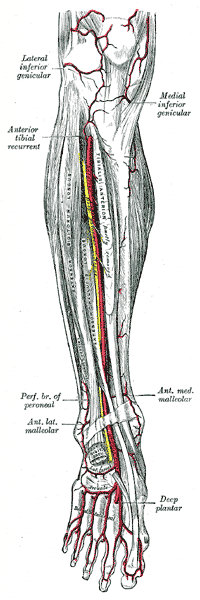
In human anatomy, the dorsalis pedis artery is a blood vessel of the lower limb. It arises from the anterior tibial artery, and ends at the first intermetatarsal space. It carries oxygenated blood to the dorsal side of the foot. It is useful for taking a pulse. It is also at risk during anaesthesia of the deep peroneal nerve.

In human anatomy, the dorsal interossei of the foot are four muscles situated between the metatarsal bones.

The extensor digitorum longus is a pennate muscle, situated at the lateral part of the front of the leg.

The extensor digitorum brevis muscle is a muscle on the upper surface of the foot that helps extend digits 2 through 4.

In human anatomy, plantar interossei muscles are three muscles located between the metatarsal bones in the foot.

The deep fibular nerve begins at the bifurcation of the common fibular nerve between the fibula and upper part of the fibularis longus, passes infero-medially, deep to the extensor digitorum longus, to the anterior surface of the interosseous membrane, and comes into relation with the anterior tibial artery above the middle of the leg; it then descends with the artery to the front of the ankle-joint, where it divides into a lateral and a medial terminal branch.

The medial plantar nerve is the larger of the two terminal divisions of the tibial nerve, which accompanies the medial plantar artery.

The lateral plantar artery, much larger than the medial, passes obliquely lateralward and forward to the base of the fifth metatarsal bone.
The plantar metatarsal arteries are four in number, arising from the convexity of the plantar arch. They run forward between the metatarsal bones and in contact with the Interossei. They are located in the fourth layer of the foot.
The medial plantar artery, much smaller than the lateral plantar artery, passes forward along the medial side of the foot.
The deep plantar artery descends into the sole of the foot, between the two heads of the 1st interosseous dorsalis, and unites with the termination of the lateral plantar artery, to complete the plantar arch.
The arcuate artery of the foot arises from dorsalis pedis slightly anterior to the lateral tarsal artery, specifically over the naviculocuneiform joint; it passes lateralward, over the bases of the lateral four metatarsal bones, beneath the tendons of the extensor digitorum brevis, its direction being influenced by its point of origin; and it terminates in the lateral tarsal artery. It communicates with the plantar arteries through the perforating arteries of the foot.
The plantar arch is a circulatory anastomosis formed from:
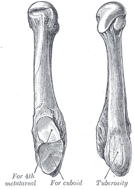
The fifth metatarsal bone is a long bone in the foot, and is palpable along the distal outer edges of the feet. It is the second smallest of the five metatarsal bones. The fifth metatarsal is analogous to the fifth metacarpal bone in the hand.

The fourth metatarsal bone is a long bone in the foot. It is smaller in size than the third metatarsal bone and is the third longest of the five metatarsal bones. The fourth metatarsal is analogous to the fourth metacarpal bone in the hand
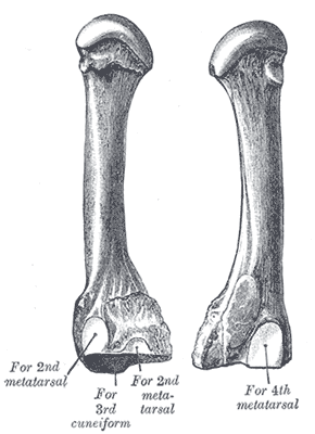
The third metatarsal bone is a long bone in the foot. It is the second longest metatarsal. The longest being the second metatarsal. The third metatarsal is analogous to the third metacarpal bone in the hand

The second metatarsal bone is a long bone in the foot. It is the longest of the metatarsal bones, being prolonged backward and held firmly into the recess formed by the three cuneiform bones. The second metatarsal forms joints with the second proximal phalanx through the metatarsophalangeal joint, the cuneiform bones, third metatarsal and occasionally the first metatarsal bone.

The first metatarsal bone is the bone in the foot just behind the big toe. The first metatarsal bone is the shortest of the metatarsal bones and by far the thickest and strongest of them.
The arcuate artery of the foot gives off the second, third, and fourth dorsal metatarsal arteries, which run forward upon the corresponding Interossei dorsales; in the clefts between the toes, each divides into two dorsal digital branches for the adjoining toes.