
The tibia, also known as the shinbone or shankbone, is the larger, stronger, and anterior (frontal) of the two bones in the leg below the knee in vertebrates ; it connects the knee with the ankle. The tibia is found on the medial side of the leg next to the fibula and closer to the median plane. The tibia is connected to the fibula by the interosseous membrane of leg, forming a type of fibrous joint called a syndesmosis with very little movement. The tibia is named for the flute tibia. It is the second largest bone in the human body, after the femur. The leg bones are the strongest long bones as they support the rest of the body.

The fibula or calf bone is a leg bone on the lateral side of the tibia, to which it is connected above and below. It is the smaller of the two bones and, in proportion to its length, the most slender of all the long bones. Its upper extremity is small, placed toward the back of the head of the tibia, below the knee joint and excluded from the formation of this joint. Its lower extremity inclines a little forward, so as to be on a plane anterior to that of the upper end; it projects below the tibia and forms the lateral part of the ankle joint.

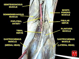
The popliteal artery is a deeply placed continuation of the femoral artery opening in the distal portion of the adductor magnus muscle. It courses through the popliteal fossa and ends at the lower border of the popliteus muscle, where it branches into the anterior and posterior tibial arteries.

The popliteal vein is a vein of the lower limb. It is formed from the anterior tibial vein and the posterior tibial vein. It travels medial to the popliteal artery, and becomes the femoral vein. It drains blood from the leg. It can be assessed using medical ultrasound. It can be affected by popliteal vein entrapment.
The anterior tibial vein is a vein in the lower leg.
In anatomy, the fibular veins are accompanying veins of the fibular artery.
The posterior tibial artery of the lower limb is an artery that carries blood to the posterior compartment of the leg and plantar surface of the foot. It branches from the popliteal artery via the tibial-fibular trunk.
The anterior tibial artery is an artery of the leg. It carries blood to the anterior compartment of the leg and dorsal surface of the foot, from the popliteal artery.

The tibial nerve is a branch of the sciatic nerve. The tibial nerve passes through the popliteal fossa to pass below the arch of soleus.
The biceps femoris is a muscle of the thigh located to the posterior, or back. As its name implies, it consists of two heads; the long head is considered part of the hamstring muscle group, while the short head is sometimes excluded from this characterization, as it only causes knee flexion and is activated by a separate nerve.
The common fibular nerve is a nerve in the lower leg that provides sensation over the posterolateral part of the leg and the knee joint. It divides at the knee into two terminal branches: the superficial fibular nerve and deep fibular nerve, which innervate the muscles of the lateral and anterior compartments of the leg respectively. When the common fibular nerve is damaged or compressed, foot drop can ensue.

The popliteal fossa is a shallow depression located at the back of the knee joint. The bones of the popliteal fossa are the femur and the tibia. Like other flexion surfaces of large joints, it is an area where blood vessels and nerves pass relatively superficially, and with an increased number of lymph nodes.
The posterior tibial recurrent artery, an inconstant branch, is given off from the anterior tibial before that vessel passes through the gap between superior tibio-fibular joint and upper border of interosseous membrane.

The interosseous membrane of the leg extends between the interosseous crests of the tibia and fibula, helps stabilize the Tib-Fib relationship and separates the muscles on the front from those on the back of the leg.
The popliteal artery entrapment syndrome (PAES) is an uncommon pathology that occurs when the popliteal artery is compressed by the surrounding popliteal fossa myofascial structures. This results in claudication and chronic leg ischemia. This condition mainly occurs more in young athletes than in the elderlies. Elderlies, who present with similar symptoms, are more likely to be diagnosed with peripheral artery disease with associated atherosclerosis. Patients with PAES mainly present with intermittent feet and calf pain associated with exercises and relieved with rest. PAES can be diagnosed with a combination of medical history, physical examination, and advanced imaging modalities such as duplex ultrasound, computer tomography, or magnetic resonance angiography. Management can range from non-intervention to open surgical decompression with a generally good prognosis. Complications of untreated PAES can include stenotic artery degeneration, complete popliteal artery occlusion, distal arterial thromboembolism, or even formation of an aneurysm.

The posterior compartment of the thigh is one of the fascial compartments that contains the knee flexors and hip extensors known as the hamstring muscles, as well as vascular and nervous elements, particularly the sciatic nerve.
The circumflex fibular artery is a branch of the posterior tibial artery which supplies blood to the knee.

The following outline is provided as an overview of and topical guide to human anatomy:
In anatomy, the fibular artery, also known as the peroneal artery, supplies blood to the lateral compartment of the leg. It arises from the tibial-fibular trunk.
