Related Research Articles
A testicle or testis is the male gonad in all bilaterians, including humans. It is homologous to the female ovary. The functions of the testicles are to produce both sperm and androgens, primarily testosterone. Testosterone release is controlled by the anterior pituitary luteinizing hormone, whereas sperm production is controlled both by the anterior pituitary follicle-stimulating hormone and gonadal testosterone.

The femoral artery is a large artery in the thigh and the main arterial supply to the thigh and leg. The femoral artery gives off the deep femoral artery and descends along the anteromedial part of the thigh in the femoral triangle. It enters and passes through the adductor canal, and becomes the popliteal artery as it passes through the adductor hiatus in the adductor magnus near the junction of the middle and distal thirds of the thigh.

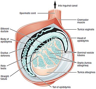
The spermatic cord is the cord-like structure in males formed by the vas deferens and surrounding tissue that runs from the deep inguinal ring down to each testicle. Its serosal covering, the tunica vaginalis, is an extension of the peritoneum that passes through the transversalis fascia. Each testicle develops in the lower thoracic and upper lumbar region and migrates into the scrotum. During its descent it carries along with it the vas deferens, its vessels, nerves etc. There is one on each side.

The inguinal canal is a passage in the anterior abdominal wall on each side of the body, which in males, convey the spermatic cords and in females, the round ligament of the uterus. The inguinal canals are larger and more prominent in males.

The male reproductive system consists of a number of sex organs that play a role in the process of human reproduction. These organs are located on the outside of the body, and within the pelvis.

The round ligament of the uterus is a ligament that connects the uterus to the labia majora. It originates at the junction of the uterus and uterine tube. It passes through the inguinal canal to insert at the labium majus.

The ovarian artery is an artery that supplies oxygenated blood to the ovary in females. It arises from the abdominal aorta below the renal artery. It can be found within the suspensory ligament of the ovary, anterior to the ovarian vein and ureter.

The membranous layer of the superficial fascia of the perineum is the deeper layer of the superficial perineal fascia. It is thin, aponeurotic in structure, and of considerable strength, serving to bind down the muscles of the root of the penis. Colles' fascia emerges from the perineal membrane, which divides the base of the penis from the prostate. Colles' fascia emerges from the inferior side of the perineal membrane and continues along the ventral (inferior) penis without covering the scrotum. It separates the skin and subcutaneous fat from the superficial perineal pouch.

The posterior scrotal branches are two in number, medial and lateral. They are branches of the perineal nerve, which is itself a branch of the pudendal nerve. The pudendal nerve arises from spinal roots S2 through S4, travels through the pudendal canal on the fascia of the obturator internus muscle, and gives off the perineal nerve in the perineum. The major branch of the perineal nerve is the posterior scrotal/posterior labial.
In anatomy, arterial tree is used to refer to all arteries and/or the branching pattern of the arteries. This article regards the human arterial tree. Starting from the aorta:

The superficial external pudendal artery is one of the three pudendal arteries. It arises from the medial side of the femoral artery, close to the superficial epigastric artery and superficial iliac circumflex artery.

The superficial perineal pouch is a compartment of the perineum.

The urogenital triangle is the anterior part of the perineum. In female mammals, it contains the vagina and associated parts of the internal genitalia.

The posterior labial nerves are branches of the pudendal nerve. They supply the female labia majora.

Related to the urinary bladder, anteriorly there are the following folds:
The posterior scrotal arteries are branches of the internal pudendal artery.

The following outline is provided as an overview of and topical guide to human anatomy:
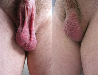
In most terrestrial mammals, the scrotum or scrotal sac is a part of the external male genitalia located at the base of the penis. It consists of a sac of skin containing the external spermatic fascia, testicles, epididymides, and vasa deferentia. The scrotum will usually tighten during penile erection and when exposed to cold temperatures.

The septum of scrotum or scrotal septum is an incomplete vertical wall (septum) that divides the scrotum into two compartments –each containing a single testis. It consists of flexible connective tissue and nonstriated muscle. The site of the median septum is apparent on the surface of the scrotum along a median longitudinal ridge called the scrotal raphe. The perineal raphe further extends forward to the undersurface of the penis and backward to the anal opening. The purpose of the median septum is to compartmentalize each testis in order to prevent friction or trauma.
Scrotal arteries may refer to:
References
- ↑ Zhao Yue-Qiang; Zhang Jie; Yu Mo-Sheng; Long Dao-Chou (2009-11-01). "Functional Restoration of Penis With Partial Defect by Scrotal Skin Flap". Journal of Urology. 182 (5): 2358–2361. doi:10.1016/j.juro.2009.07.048. PMID 19762052.
| | This cardiovascular system article is a stub. You can help Wikipedia by expanding it. |