
The human leg, in the general word sense, is the entire lower limb of the human body, including the foot, thigh or sometimes even the hip or gluteal region. However, the definition in human anatomy refers only to the section of the lower limb extending from the knee to the ankle, also known as the crus or, especially in non-technical use, the shank. Legs are used for standing, and all forms of locomotion including recreational such as dancing, and constitute a significant portion of a person's mass. Female legs generally have greater hip anteversion and tibiofemoral angles, but shorter femur and tibial lengths than those in males.

The femoral artery is a large artery in the thigh and the main arterial supply to the thigh and leg. The femoral artery gives off the deep femoral artery and descends along the anteromedial part of the thigh in the femoral triangle. It enters and passes through the adductor canal, and becomes the popliteal artery as it passes through the adductor hiatus in the adductor magnus near the junction of the middle and distal thirds of the thigh.

The femoral triangle is an anatomical region of the upper third of the thigh. It is a subfascial space which appears as a triangular depression below the inguinal ligament when the thigh is flexed, abducted and laterally rotated.

The popliteal artery is a deeply placed continuation of the femoral artery opening in the distal portion of the adductor magnus muscle. It courses through the popliteal fossa and ends at the lower border of the popliteus muscle, where it branches into the anterior and posterior tibial arteries.

The tibial nerve is a branch of the sciatic nerve. The tibial nerve passes through the popliteal fossa to pass below the arch of soleus.

The adductor magnus is a large triangular muscle, situated on the medial side of the thigh.

The superior genicular arteries, two in number, arise one on either side of the popliteal artery, and wind around the femur immediately above its condyles to the front of the knee-joint. The medial superior genicular artery is on the inside of the knee and the lateral superior genicular artery is on the outside.

The lateral superior genicular artery is a branch of the popliteal artery that supplies a portion of the knee joint.
The medial superior genicular, a branch of the popliteal artery, runs in front of the Semimembranosus and Semitendinosus, above the medial head of the Gastrocnemius, and passes beneath the tendon of the Adductor magnus.
The inferior genicular arteries, two in number, arise from the popliteal beneath the gastrocnemius. On the inside of the knee, is the medial inferior genicular artery, and on the outer side is the lateral inferior genicular artery.
The medial inferior genicular is an artery of the leg.

The lateral circumflex femoral artery is an artery in the upper thigh. It is usually a branch of the profunda femoris artery, and produces three branches. It is mostly distributed to the muscles of the lateral thigh, supplying arterial blood to muscles of the knee extensor group.

The adductor canal is an aponeurotic tunnel in the middle third of the thigh giving passage to parts of the femoral artery, vein, and nerve. It extends from the apex of the femoral triangle to the adductor hiatus.
In anatomy, arterial tree is used to refer to all arteries and/or the branching pattern of the arteries. This article regards the human arterial tree. Starting from the aorta:
The saphenous nerve is the largest cutaneous branch of the femoral nerve. It is derived from the lumbar plexus (L3-L4). It is a strictly sensory nerve, and has no motor function. It commences in the proximal (upper) thigh and travels along the adductor canal. Upon exiting the adductor canal, the saphenous nerve terminates by splitting into two terminal branches: the sartorial nerve, and the infrapatellar nerve. The saphenous nerve is responsible for providing sensory innervation to the skin of the anteromedial leg.

In human anatomy, the adductor hiatus also known as hiatus magnus is a hiatus (gap) between the adductor magnus muscle and the femur that allows the passage of the femoral vessels from the anterior thigh to the posterior thigh and then the popliteal fossa. It is the termination of the adductor canal and lies about 8–13.5 cm. superior to the adductor tubercle.
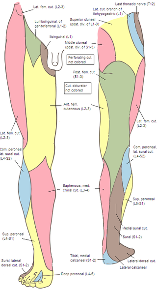
The anterior cutaneous branches of the femoral nerve consist of the following nerves: intermediate cutaneous nerve and medial cutaneous nerve.
The patellar network is an intricate network of blood vessels around and above the patella, and on the contiguous ends of the femur and tibia, forming a superficial and a deep plexus.
The subsartorial plexus is a plexus of nerves that is located under the sartorius muscle.
Genicular artery may refer to one of six arteries in the human leg, most of which anastomose in the knee region.
