
The leg is the entire lower limb of the human body, including the foot, thigh or sometimes even the hip or buttock region. The major bones of the leg are the femur, tibia, and adjacent fibula. The thigh is between the hip and knee, while the calf (rear) and shin (front) are between the knee and foot.

The tibial nerve is a branch of the sciatic nerve. The tibial nerve passes through the popliteal fossa to pass below the arch of soleus.

The superficial fibular nerve is a mixed nerve that provides motor innervation to the fibularis longus and fibularis brevis muscles, and sensory innervation to skin over the antero-lateral aspect of the leg along with the greater part of the dorsum of the foot.

The medial plantar nerve is the larger of the two terminal divisions of the tibial nerve, which accompanies the medial plantar artery.
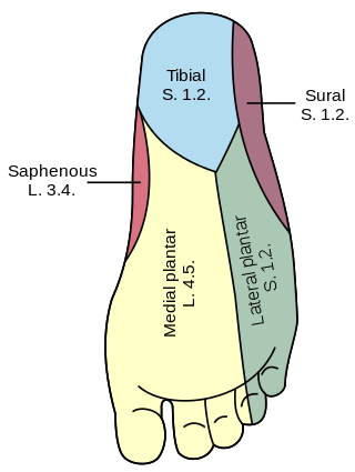
The lateral plantar nerve is a branch of the tibial nerve, in turn a branch of the sciatic nerve and supplies the skin of the fifth toe and lateral half of the fourth, as well as most of the deep muscles, its distribution being similar to that of the ulnar nerve in the hand.

The medial plantar artery, much smaller than the lateral plantar artery, passes forward along the medial side of the foot.

In the palm of the hand the median nerve is covered by the skin and the palmar aponeurosis, and rests on the tendons of the flexor muscles. Immediately after emerging from under the transverse carpal ligament the median nerve becomes enlarged and flattened and splits into a smaller, lateral, and a larger, medial portion.

The proper palmar digital arteries travel along the sides of the phalanges, each artery lying just below its corresponding digital nerve.
A neurovascular bundle is a structure that binds nerves and veins with connective tissue so that they travel in tandem through the body.

The following outline is provided as an overview of and topical guide to human anatomy:
The superficial branch of the lateral plantar nerve splits into a proper and a common plantar digital nerve:

Dorsal digital nerves of foot are branches of the intermediate dorsal cutaneous nerve, medial dorsal cutaneous nerve, sural nerve and deep fibular nerve.
The proper plantar digital nerves of lateral plantar nerve are nerves of the foot that arise from the superficial branch of the lateral plantar nerve. The superficial branch splits into a proper digital nerve and a common digital nerve:
The common plantar digital nerves of lateral plantar nerve are nerves of the foot. The common digital nerve communicates with the third common digital branch of the medial plantar nerve and divides into two proper digital nerves which supply the adjoining sides of the fourth and fifth toes.
The proper plantar digital nerves of medial plantar nerve are nerves of the foot. They primarily arise from the medial plantar nerve's superficial and deep branches. The superficial branch of the medial plantar nerve turns into a proper digital nerve and is responsible for supplies the medial side of the great toe.
The common plantar digital nerves of medial plantar nerve are nerves of the foot. The three common digital nerves pass between the divisions of the plantar aponeurosis, and each splits into two proper digital nerves:
The common plantar digital arteries are arteries of the foot.
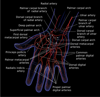
Dorsal digital arteries arise from the bifurcation of dorsal metacarpal arteries. They travel along the sides and dorsal aspects of the phalanges of the middle finger, ring finger, and little finger. They communicate with the proper palmar digital arteries.
Plantar digital nerves may refer to;
This page is based on this
Wikipedia article Text is available under the
CC BY-SA 4.0 license; additional terms may apply.
Images, videos and audio are available under their respective licenses.
