Related Research Articles

The femoral artery is a large artery in the thigh and the main arterial supply to the thigh and leg. The femoral artery gives off the deep femoral artery and descends along the anteromedial part of the thigh in the femoral triangle. It enters and passes through the adductor canal, and becomes the popliteal artery as it passes through the adductor hiatus in the adductor magnus near the junction of the middle and distal thirds of the thigh.

The femoral triangle is an anatomical region of the upper third of the thigh. It is a subfascial space which appears as a triangular depression below the inguinal ligament when the thigh is flexed, abducted and laterally rotated.

The deep femoral artery also known as the deep artery of the thigh, or profunda femoris artery, is a large branch of the femoral artery. It travels more deeply ("profoundly") than the rest of the femoral artery. It gives rise to the lateral circumflex femoral artery and medial circumflex femoral artery, and the perforating arteries, terminating within the thigh.

The popliteal artery is a deeply placed continuation of the femoral artery opening in the distal portion of the adductor magnus muscle. It courses through the popliteal fossa and ends at the lower border of the popliteus muscle, where it branches into the anterior and posterior tibial arteries.

The adductor magnus is a large triangular muscle, situated on the medial side of the thigh.

The internal iliac artery is the main artery of the pelvis.

The iliolumbar artery is the first branch of the posterior trunk of the internal iliac artery.

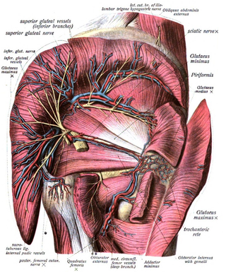
The superior gluteal artery is the terminal branch of the posterior division of the internal iliac artery. It exits the pelvis through the greater sciatic foramen before splitting into a superficial branch and a deep branch.
The inferior gluteal artery is a terminal branch of the anterior trunk of the internal iliac artery. It exits the pelvis through the greater sciatic foramen. It is distributed chiefly to the buttock and the back of the thigh.

The obturator artery is a branch of the internal iliac artery that passes antero-inferiorly on the lateral wall of the pelvis, to the upper part of the obturator foramen, and, escaping from the pelvic cavity through the obturator canal, it divides into an anterior branch and a posterior branch.
The inferior gluteal veins are venae comitantes of the inferior gluteal artery. They commence in the superior/proximal posterior thigh. They enter the pelvis through the lower part of the greater sciatic foramen. They converge to form a single vessel before emptying into the into the distal portion of the internal iliac vein.

The lateral superior genicular artery is a branch of the popliteal artery that supplies a portion of the knee joint.

The lateral circumflex femoral artery is an artery in the upper thigh. It is usually a branch of the profunda femoris artery, and produces three branches. It is mostly distributed to the muscles of the lateral thigh, supplying arterial blood to muscles of the knee extensor group.
In anatomy, arterial tree is used to refer to all arteries and/or the branching pattern of the arteries. This article regards the human arterial tree. Starting from the aorta:

The deep circumflex iliac artery is an artery in the pelvis that travels along the iliac crest of the pelvic bone.

The perforating arteries are branches of the deep artery of the thigh, usually three in number, so named because they perforate the tendon of the adductor magnus to reach the back of the thigh. They pass backward near the linea aspera of the femur underneath the small tendinous arches of the adductor magnus muscle.
The patellar network is an intricate network of blood vessels around and above the patella, and on the contiguous ends of the femur and tibia, forming a superficial and a deep plexus.
The superficial branch of medial circumflex femoral artery appears between the quadratus femoris and upper border of the adductor magnus, and anastomoses with the inferior gluteal artery, lateral femoral circumflex artery, and first of the perforating arteries of the deep femoral artery.
The trochanteric anastomosis provides circulation around the head of the femur. It includes the superior gluteal artery and the medial and lateral circumflex femoral arteries.
The genicular arteries are six arteries in the human leg, five of which are branches of the popliteal artery, that anastomose in the knee region in the patellar network or genicular anastomosis. They supply blood to the patella, together with contributions from the descending genicular artery, anterior tibial recurrent artery, and descending branch of lateral circumflex femoral artery.
References
![]() This article incorporates text in the public domain from page 620 of the 20th edition of Gray's Anatomy (1918)
This article incorporates text in the public domain from page 620 of the 20th edition of Gray's Anatomy (1918)