
The cervix or cervix uteri is the lower part of the uterus in the human female reproductive system. The cervix is usually 2 to 3 cm long and roughly cylindrical in shape, which changes during pregnancy. The narrow, central cervical canal runs along its entire length, connecting the uterine cavity and the lumen of the vagina. The opening into the uterus is called the internal os, and the opening into the vagina is called the external os. The lower part of the cervix, known as the vaginal portion of the cervix, bulges into the top of the vagina. The cervix has been documented anatomically since at least the time of Hippocrates, over 2,000 years ago.

The uterus or womb is a major female hormone-responsive secondary sex organ of the reproductive system in humans and most other mammals. In the human, the lower end of the uterus, the cervix, opens into the vagina, while the upper end, the fundus, is connected to the fallopian tubes. It is within the uterus that the fetus develops during gestation. In the human embryo, the uterus develops from the paramesonephric ducts which fuse into the single organ known as a simplex uterus. The uterus has different forms in many other animals and in some it exists as two separate uteri known as a duplex uterus.
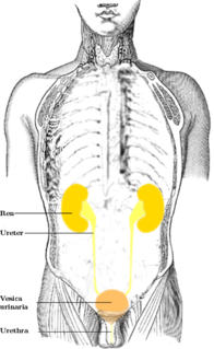
In human anatomy, the ureters are tubes made of smooth muscle fibers that propel urine from the kidneys to the urinary bladder. In the adult, the ureters are usually 25–30 cm (10–12 in) long and around 3–4 mm (0.12–0.16 in) in diameter. Histologically, the ureter is lined by the urothelium, a type of transitional epithelium, and has an additional smooth muscle layer in the more distal one-third to assist with peristalsis.

The female reproductive system is made up of the internal and external sex organs that function in reproduction of new offspring. In the human the female reproductive system is immature at birth and develops to maturity at puberty to be able to produce gametes, and to carry a foetus to full term. The internal sex organs are the uterus, Fallopian tubes, and ovaries. The uterus or womb accommodates the embryo which develops into the foetus. The uterus also produces vaginal and uterine secretions which help the transit of sperm to the Fallopian tubes. The ovaries produce the ova. The external sex organs are also known as the genitals and these are the organs of the vulva including the labia, clitoris, and vaginal opening. The vagina is connected to the uterus at the cervix.
Vaginal bleeding is any bleeding through the vagina, including bleeding from the vaginal wall itself, as well as bleeding from another location of the female reproductive system, often the uterus. Generally, it is either part of a normal menstrual cycle or is caused by hormonal or other problems of the reproductive system, such as abnormal uterine bleeding. Vaginal bleeding may occur at any age, but always needs investigation when encountered in children or in postmenopausal women. Vaginal bleeding during pregnancy may indicate a possible pregnancy complication that needs to be medically addressed.

The recto-uterine pouch, also known by various other names, is the extension of the peritoneal cavity between the rectum and the posterior wall of the uterus in the female human body.

Uterine artery embolization is a procedure where an interventional radiologist uses a catheter to deliver small particles that block the blood supply to the uterine body. The procedure is done for the treatment of uterine fibroids and adenomyosis. Given that this minimally invasive procedure is commonly used in the treatment of uterine fibroids it is also called uterine fibroid embolization (UFE).

The sacrospinous ligament is a thin, triangular ligament in the human pelvis. The base of the ligament is attached to the outer edge of the sacrum and coccyx, and the tip of the ligament attaches to the spine of the ischium, a bone in the human pelvis. Its fibres are intermingled with the sacrotuberous ligament.

The round ligament of the uterus originates at the uterine horns, in the parametrium. The round ligament exits the pelvis via the deep inguinal ring, passes through the inguinal canal and continues on to the labia majora where its fibers spread and mix with the tissue of the mons pubis.
The vaginal artery is an artery in females that supplies blood to the vagina and the base of the bladder.

The inferior vesical artery or inferior vesicle artery is an artery in the pelvis that supplies the lower part of the bladder.
The ovarian artery is an artery that supplies oxygenated blood to the ovary in females. It arises from the abdominal aorta below the renal artery. It can be found in the suspensory ligament of the ovary, anterior to the ovarian vein and ureter.

The cervical canal is the spindle-shaped, flattened canal of the cervix, the neck of the uterus.
The Uterovaginal plexus is a division of the inferior hypogastric plexus. In older texts, it is referred to as two structures, the "vaginal plexus" and "uterine plexus".

The fornices of the vagina are the superior portions of the vagina, extending into the recesses created by the vaginal portion of cervix. The word "fornix" is Latin for "arch".

The vaginal venous plexuses are placed at the sides of the vagina; they communicate with the uterine venous plexuses, vesical venous plexus, and rectal venous plexuses, and are drained by the vaginal veins, one on either side, into the hypogastric veins.
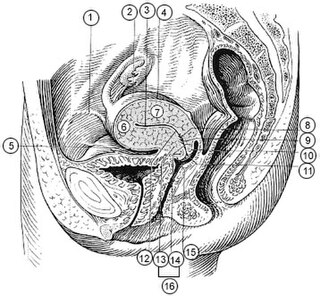
The supravaginal portion of the cervix is separated in front from the bladder by fibrous tissue (parametrium), which extends also on to its sides and lateralward between the layers of the broad ligaments.
A vaginal disease is a pathological condition that affects part or all of the vagina.
The public domain consists of all the creative works to which no exclusive intellectual property rights apply. Those rights may have expired, been forfeited, expressly waived, or may be inapplicable.

Gray's Anatomy is an English language textbook of human anatomy originally written by Henry Gray and illustrated by Henry Vandyke Carter. Earlier editions were called Anatomy: Descriptive and Surgical, Anatomy of the Human Body and Gray's Anatomy: Descriptive and Applied, but the book's name is commonly shortened to, and later editions are titled, Gray's Anatomy. The book is widely regarded as an extremely influential work on the subject, and has continued to be revised and republished from its initial publication in 1858 to the present day. The latest edition of the book, the 41st, was published in September 2015.

