The thoracic diaphragm, or simply the diaphragm, is a sheet of internal skeletal muscle in humans and other mammals that extends across the bottom of the thoracic cavity. The diaphragm is the most important muscle of respiration, and separates the thoracic cavity, containing the heart and lungs, from the abdominal cavity: as the diaphragm contracts, the volume of the thoracic cavity increases, creating a negative pressure there, which draws air into the lungs. Its high oxygen consumption is noted by the many mitochondria and capillaries present; more than in any other skeletal muscle.

The rectus abdominis muscle, also known as the "abdominal muscle" or simply the "abs", is a pair of segmented skeletal muscle on the ventral aspect of a person's abdomen. The paired muscle is separated at the midline by a band of dense connective tissue called the linea alba, and the connective tissue defining each lateral margin of the rectus abdominus is the linea semilunaris. The muscle extends from the pubic symphysis, pubic crest and pubic tubercle inferiorly, to the xiphoid process and costal cartilages of the 5th–7th ribs superiorly.

The renal arteries are paired arteries that supply the kidneys with blood. Each is directed across the crus of the diaphragm, so as to form nearly a right angle.

In human anatomy, the superior mesenteric artery (SMA) is an artery which arises from the anterior surface of the abdominal aorta, just inferior to the origin of the celiac trunk, and supplies blood to the intestine from the lower part of the duodenum through two-thirds of the transverse colon, as well as the pancreas.

The transverse abdominal muscle (TVA), also known as the transverse abdominis, transversalis muscle and transversus abdominis muscle, is a muscle layer of the anterior and lateral abdominal wall, deep to the internal oblique muscle. It is thought by most fitness instructors to be a significant component of the core.

In human anatomy, the inferior mesenteric artery (IMA) is the third main branch of the abdominal aorta and arises at the level of L3, supplying the large intestine from the distal transverse colon to the upper part of the anal canal. The regions supplied by the IMA are the descending colon, the sigmoid colon, and part of the rectum.

The external iliac arteries are two major arteries which bifurcate off the common iliac arteries anterior to the sacroiliac joint of the pelvis.
The abdominal external oblique muscle is the largest and outermost of the three flat abdominal muscles of the lateral anterior abdomen.

The quadratus lumborum muscle, informally called the QL, is a paired muscle of the left and right posterior abdominal wall. It is the deepest abdominal muscle, and commonly referred to as a back muscle. Each is irregular and quadrilateral in shape.
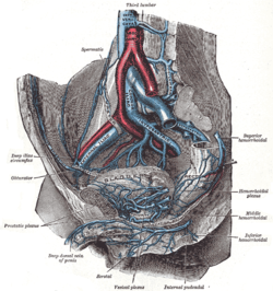
The internal iliac artery is the main artery of the pelvis.

In human anatomy, the inferior epigastric artery is an artery that arises from the external iliac artery. It is accompanied by the inferior epigastric vein; inferiorly, these two inferior epigastric vessels together travel within the lateral umbilical fold The inferior epigastric artery then traverses the arcuate line of rectus sheath to enter the rectus sheath, then anastomoses with the superior epigastric artery within the rectus sheath.

The iliohypogastric nerve is a nerve that originates from the lumbar plexus that supplies sensation to skin over the lateral gluteal and hypogastric regions and motor to the internal oblique muscles and transverse abdominal muscles.
The ilioinguinal nerve is a branch of the first lumbar nerve (L1). It separates from the first lumbar nerve along with the larger iliohypogastric nerve. It emerges from the lateral border of the psoas major just inferior to the iliohypogastric, and passes obliquely across the quadratus lumborum and iliacus. The ilioinguinal nerve then perforates the transversus abdominis near the anterior part of the iliac crest, and communicates with the iliohypogastric nerve between the transversus and the internal oblique muscle.
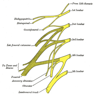
The lumbar nerves are the five pairs of spinal nerves emerging from the lumbar vertebrae. They are divided into posterior and anterior divisions.

The inferior phrenic artery is a bilaterally paired artery of the abdominal cavity which represents the main source of arterial supply to the diaphragm. Each artery usually arises either from the coeliac trunk or the abdominal aorta, however, their origin is highly variable and the different sites of origin are different for the left artery and right artery. The superior suprarenal artery is a branch of the inferior phrenic artery.
The median sacral artery is a small artery that arises posterior to the abdominal aorta and superior to its bifurcation.

The inferior thyroid artery is an artery in the neck. It arises from the thyrocervical trunk and passes upward, in front of the vertebral artery and longus colli muscle. It then turns medially behind the carotid sheath and its contents, and also behind the sympathetic trunk, the middle cervical ganglion resting upon the vessel.
The subcostal nerve is a mixed motor and sensory nerve contributing to the lumbar plexus. It runs along the lower border of the twelfth rib, often gives a communicating branch to the first lumbar nerve, and passes under the lateral lumbocostal arch.
The subcostal arteries, so named because they lie below the last ribs, constitute the lowest pair of branches derived from the thoracic aorta, and are in series with the intercostal arteries.

The intercostal arteries are a group of arteries passing within an intercostal space. There are 9 anterior and 11 posterior intercostal arteries on each side of the body. The anterior intercostal arteries are branches of the internal thoracic artery and its terminal branch - the musculophrenic artery. The posterior intercostal arteries are branches of the supreme intercostal artery and thoracic aorta.