
The levator ani is a broad, thin muscle group, situated on either side of the pelvis. It is formed from three muscle components: the pubococcygeus, the iliococcygeus, and the puborectalis.

The sigmoid colon is the part of the large intestine that is closest to the rectum and anus. It forms a loop that averages about 35–40 centimetres (14–16 in) in length. The loop is typically shaped like a Greek letter sigma (ς) or Latin letter S. This part of the colon normally lies within the pelvis, but due to its freedom of movement it is liable to be displaced into the abdominal cavity.

The anal canal is the part that connects the rectum to the anus, located below the level of the pelvic diaphragm. It is located within the anal triangle of the perineum, between the right and left ischioanal fossa. As the final functional segment of the bowel, it functions to regulate release of excrement by two muscular sphincter complexes. The anus is the aperture at the terminal portion of the anal canal.

In human anatomy, the inferior mesenteric artery (IMA) is the third main branch of the abdominal aorta and arises at the level of L3, supplying the large intestine from the distal transverse colon to the upper part of the anal canal. The regions supplied by the IMA are the descending colon, the sigmoid colon, and part of the rectum.

The internal iliac artery is the main artery of the pelvis.

In human anatomy, the inferior epigastric artery is an artery that arises from the external iliac artery. It is accompanied by the inferior epigastric vein; inferiorly, these two inferior epigastric vessels together travel within the lateral umbilical fold The inferior epigastric artery then traverses the arcuate line of rectus sheath to enter the rectus sheath, then anastomoses with the superior epigastric artery within the rectus sheath.

The uterine artery is an artery that supplies blood to the uterus in females.

The iliolumbar artery is the first branch of the posterior trunk of the internal iliac artery.

The middle rectal artery is an artery in the pelvis that supplies blood to the rectum.

The inferior rectal artery is an artery that supplies blood to the lower third of the anal canal below the pectinate line.
The sigmoid arteries are 2–5 branches of the inferior mesenteric artery that are distributed to the distal descending colon and the sigmoid colon.

The right colic artery is an artery of the abdomen, a branch of the superior mesenteric artery supplying the ascending colon. It divides into two terminal branches - an ascending branch and a descending branch - which form anastomoses with the middle colic artery, and ileocolic artery (respectively).

The middle colic artery is an artery of the abdomen; a branch of the superior mesenteric artery distributed to parts of the ascending and transverse colon. It usually divides into two terminal branches - a left one and a right one - which go on to form anastomoses with the left colic artery, and right colic artery (respectively), thus participating in the formation of the marginal artery of the colon.
The left colic artery is a branch of the inferior mesenteric artery distributed to the descending colon, and left part of the transverse colon. It ends by dividing into an ascending branch and a descending branch; the terminal branches of the two branches go on to form anastomoses with the middle colic artery, and a sigmoid artery (respectively).

The ileocolic artery is the lowest branch arising from the concavity of the superior mesenteric artery. It supplies the cecum, ileum, and appendix.

The inferior mesenteric plexus is derived chiefly from the aortic plexus.
The inferior rectal nerves usually branch from the pudendal nerve but occasionally arises directly from the sacral plexus; they cross the ischiorectal fossa along with the inferior rectal artery and veins, toward the anal canal and the lower end of the rectum, and is distributed to the sphincter ani externus and to the integument (skin) around the anus.
The inferior mesenteric vein begins in the rectum as the superior rectal vein, which has its origin in the hemorrhoidal plexus, and through this plexus communicates with the middle and inferior hemorrhoidal veins.

The inferior mesenteric lymph nodes consist of:

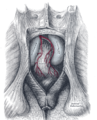
The rectum is the final straight portion of the large intestine in humans and some other mammals, and the gut in others. The adult human rectum is about 12 centimetres (4.7 in) long, and begins at the rectosigmoid junction at the level of the third sacral vertebra or the sacral promontory depending upon what definition is used. Its diameter is similar to that of the sigmoid colon at its commencement, but it is dilated near its termination, forming the rectal ampulla. It terminates at the level of the anorectal ring or the dentate line, again depending upon which definition is used. In humans, the rectum is followed by the anal canal, which is about 4 centimetres (1.6 in) long, before the gastrointestinal tract terminates at the anal verge. The word rectum comes from the Latin rēctumintestīnum, meaning straight intestine.

