Renal artery

Last updated
Renal artery
Gray1121.png
Renal arteries branching left and right from the aorta (in red)
Kidney PioM.png
Details
Source Abdominal aorta
Branches Inferior suprarenal artery, segmental arteries, Ovarian artery
Vein Renal vein
Supplies Kidneys
Identifiers
Latin arteria renalis
MeSH D012077
TA98 A12.2.12.075
TA2 4269
FMA 14751
Anatomical terminology

The renal arteries are paired arteries that supply the kidneys with blood. Each is directed across the crus of the diaphragm, so as to form nearly a right angle.

Contents

The renal arteries carry a large portion of total blood flow to the kidneys. Up to a third of total cardiac output can pass through the renal arteries to be filtered by the kidneys.

Structure

The renal arteries normally arise at a 90° angle off of the left interior side of the abdominal aorta, immediately below the superior mesenteric artery. [1] They have a radius of approximately 0.25  cm, [2] 0.26 cm at the root. [3] The measured mean diameter can differ depending on the imaging method used. For example, the diameter was found to be 5.04 ± 0.74 mm using ultrasound but 5.68 ± 1.19 mm using angiography. [4] [5]

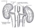
Due to the anatomical position of the aorta, the inferior vena cava, and the kidneys, the right renal artery is normally longer than the left renal artery. [1] [6]

Branches

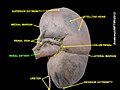
Before reaching the hilus of the kidney, each artery divides into four or five branches. The anterior branches (the upper, middle, lower and apical segmental arteries) lie between the renal vein and ureter, the vein being in front, the ureter behind. The posterior branches, which are fewer in number and include the posterior segmental artery, are usually situated behind the ureter. [7]

Each vessel gives off some small inferior suprarenal branches to the suprarenal gland, the ureter, and the surrounding cellular tissue and muscles.

One or two accessory renal arteries are frequently found, especially on the left side since they usually arise from the aorta, and may come off above (more common) or below the main artery. Instead of entering the kidney at the hilus, they usually pierce the upper or lower part of the organ.

Variation

The arterial supply of the kidneys is variable and there may be one or more renal arteries supplying each kidney. [1] It is located above the renal vein. Supernumerary renal arteries (two or more arteries to a single kidney) are the most common renovascular anomaly, occurrence ranging from 25% to 40% of kidneys. [8] Aberrant renal arteries may be present, and may complicate surgical procedures. [9]

Clinical significance

Stenosis

Renal artery stenosis, or narrowing of one or both renal arteries will lead to hypertension as the affected kidneys release renin to increase blood pressure to preserve perfusion to the kidneys. RAS is typically diagnosed with duplex ultrasonography of the renal arteries. It is treated with the use of balloon angioplasty and stents, if necessary.

Atherosclerosis

Atherosclerosis can also affect the renal arteries and can lead to poor perfusion of the kidneys leading to reduced kidney function and, possibly, renal failure

Renal artery aneurysm

A dilated renal artery measuring twice its normal size indicates a renal artery aneurysm. [4]

Trauma

A renal artery is damaged in 4% of blunt traumas and 7% of penetrating traumas to the abdomen. [10]

Additional images

Related Research Articles

<span class="mw-page-title-main">Aorta</span> Largest artery in the human body

The aorta is the main and largest artery in the human body, originating from the left ventricle of the heart, branching upwards immediately after, and extending down to the abdomen, where it splits at the aortic bifurcation into two smaller arteries. The aorta distributes oxygenated blood to all parts of the body through the systemic circulation.

<span class="mw-page-title-main">Kidney</span> Organ that filters blood and produces urine in humans

In humans, the kidneys are two reddish-brown bean-shaped blood-filtering organs that are a multilobar, multipapillary form of mammalian kidneys, usually without signs of external lobulation. They are located on the left and right in the retroperitoneal space, and in adult humans are about 12 centimetres in length. They receive blood from the paired renal arteries; blood exits into the paired renal veins. Each kidney is attached to a ureter, a tube that carries excreted urine to the bladder.

<span class="mw-page-title-main">Inferior vena cava</span> One of two veinous trunks bringing deoxygenated blood back to the heart

The inferior vena cava is a large vein that carries the deoxygenated blood from the lower and middle body into the right atrium of the heart. It is formed by the joining of the right and the left common iliac veins, usually at the level of the fifth lumbar vertebra.

<span class="mw-page-title-main">Abdominal aorta</span> Largest artery in the abdomen

In human anatomy, the abdominal aorta is the largest artery in the abdominal cavity. As part of the aorta, it is a direct continuation of the descending aorta.

<span class="mw-page-title-main">Periaortic lymph nodes</span> Human lymph nodes

The periaortic lymph nodes are a group of lymph nodes that lie in front of the lumbar vertebrae near the aorta. These lymph nodes receive drainage from the gastrointestinal tract and the abdominal organs.

<span class="mw-page-title-main">Renal vein</span> Short thick veins which return blood from the kidneys to the vena cava

The renal veins in the renal circulation, are large-calibre veins that drain blood filtered by the kidneys into the inferior vena cava. There is one renal vein draining each kidney. Each renal vein is formed by the convergence of the interlobar veins of one kidney.

<span class="mw-page-title-main">Celiac artery</span> First major branch of the abdominal aorta

The celiacartery, also known as the celiac trunk or truncus coeliacus, is the first major branch of the abdominal aorta. It is about 1.25 cm in length. Branching from the aorta at thoracic vertebra 12 (T12) in humans, it is one of three anterior/ midline branches of the abdominal aorta.

<span class="mw-page-title-main">Superior mesenteric artery</span> Artery which supplies blood to the intestines and pancreas

In human anatomy, the superior mesenteric artery (SMA) is an artery which arises from the anterior surface of the abdominal aorta, just inferior to the origin of the celiac trunk, and supplies blood to the intestine from the lower part of the duodenum through two-thirds of the transverse colon, as well as the pancreas.

<span class="mw-page-title-main">Inferior mesenteric artery</span>

In human anatomy, the inferior mesenteric artery (IMA) is the third main branch of the abdominal aorta and arises at the level of L3, supplying the large intestine from the distal transverse colon to the upper part of the anal canal. The regions supplied by the IMA are the descending colon, the sigmoid colon, and part of the rectum.

<span class="mw-page-title-main">Internal iliac artery</span> Main artery of the pelvis

The internal iliac artery is the main artery of the pelvis.

<span class="mw-page-title-main">Renal circulation</span>

The renal circulation supplies the blood to the kidneys via the renal arteries, left and right, which branch directly from the abdominal aorta. Despite their relatively small size, the kidneys receive approximately 20% of the cardiac output.

<span class="mw-page-title-main">Hepatic portal system</span> System of veins comprising the hepatic portal vein and its tributaries

In human anatomy, the hepatic portal system or portal venous system is the system of veins comprising the portal vein and its tributaries. The other portal venous systems in the body are the renal portal system, and the hypophyseal portal system.

<span class="mw-page-title-main">Inferior phrenic arteries</span>

The inferior phrenic artery is a bilaterally paired artery of the abdominal cavity which represents the main source of arterial supply to the diaphragm. Each artery usually arises either from the coeliac trunk or the abdominal aorta, however, their origin is highly variable and the different sites of origin are different for the left artery and right artery. The superior suprarenal artery is a branch of the inferior phrenic artery.

<span class="mw-page-title-main">Left colic artery</span> Artery

The left colic artery is a branch of the inferior mesenteric artery distributed to the descending colon, and left part of the transverse colon. It ends by dividing into an ascending branch and a descending branch; the terminal branches of the two branches go on to form anastomoses with the middle colic artery, and a sigmoid artery (respectively).

<span class="mw-page-title-main">Middle suprarenal arteries</span> Arteries of the abdomen

The middle suprarenal artery is a paired artery in the abdomen. It is a branch of the aorta. It supplies the adrenal gland.

<span class="mw-page-title-main">Inferior suprarenal artery</span> Artery that supplies the adrenal gland

The inferior suprarenal artery is a paired artery that supplies the adrenal gland. It usually originates at the trunk of the renal artery before its terminal division, but with many common variations. It supplies the adrenal gland parenchyma, the ureter, and the surrounding cellular tissue and muscles.

<span class="mw-page-title-main">Testicular artery</span> Branch of the abdominal aorta that supplies blood to the testicle

The testicular artery is a branch of the abdominal aorta that supplies blood to the testicle. It is a paired artery, with one for each of the testicles.

<span class="mw-page-title-main">Testicular vein</span>

The testicular vein, the male gonadal vein, carries deoxygenated blood from its corresponding testis to the inferior vena cava or one of its tributaries. It is the male equivalent of the ovarian vein, and is the venous counterpart of the testicular artery.

<span class="mw-page-title-main">Outline of human anatomy</span> Overview of and topical guide to human anatomy

The following outline is provided as an overview of and topical guide to human anatomy:

References

PD-icon.svgThis article incorporates text in the public domain from page 610 of the 20th edition of Gray's Anatomy (1918)

  1. 1 2 3 Listmann, Mishan; Tubbs, R. Shane (2020-01-01), Tubbs, R. Shane; Iwanaga, Joe; Oskouian, Rod J.; Moisi, Marc (eds.), "Chapter 19 - The Abdominal Aorta", Surgical Anatomy of the Lateral Transpsoas Approach to the Lumbar Spine, St. Louis: Elsevier, pp. 185–188, ISBN   978-0-323-67376-1 , retrieved 2021-01-13
  2. Kem, D. C.; Lyons, D. F.; Wenzl, J.; Halverstadt, D.; Yu, X. (2005). "Renin-Dependent Hypertension Caused by Nonfocal Stenotic Aberrant Renal Arteries: Proof of a New Syndrome". Hypertension. 46 (2): 380–5. doi: 10.1161/01.HYP.0000171185.25749.5b . PMID   15967872.
  3. Ottesen, Johnny T.; Danielsen, Michael, eds. (2000). Mathematical Modelling in Medicine. IOS Press. ISBN   978-4-274-90318-2.[ page needed ]
  4. 1 2 Renal Artery Aneurysm at eMedicine
  5. Aytac, Suat K.; Yigit, Hasan; Sancak, Tanzer; Ozcan, Hasan (2003). "Correlation between the diameter of the main renal artery and the presence of an accessory renal artery: sonographic and angiographic evaluation". Journal of Ultrasound in Medicine. 22 (5): 433–9, quiz 440–2. doi:10.7863/jum.2003.22.5.433. PMID   12751854. S2CID   24188140.
  6. Saldarriaga, Bladimir; Pinto, Sergio A; Ballesteros, Luis E (2008). "Morphological Expression of the Renal Artery: A Direct Anatomical Study in a Colombian Half-caste Population". International Journal of Morphology. 26 (1): 31–8. doi: 10.4067/S0717-95022008000100005 .
  7. Leslie, Stephen W.; Sajjad, Hussain (11 Aug 2021). "Anatomy, Abdomen and Pelvis, Renal Artery". National Center for Biotechnology Information. PMID   29083626 . Retrieved 15 Nov 2021.
  8. Coello-Torà, Iris; Segura-Sampedro, Juan José; Pérez-Celada, Judit; Jiménez-Morillas, Patricia; Morales-Soriano, Rafael (January 2020). "[Accessory renal artery arising from infrarenal aorta, exposed during linphadenectomy due to cytoreductive surgery and HIPEC.]". Archivos Espanoles de Urologia. 73 (1): 76–77. ISSN   0004-0614. PMID   31950928.
  9. Cerny, Joseph C.; Karsch, Daniel (1973-12-01). "Aberrant renal arteries". Urology. 2 (6): 623–626. doi:10.1016/0090-4295(73)90322-1. hdl: 2027.42/33981 . ISSN   0090-4295. PMID   4766019.
  10. Kaufman, John A. (2004-01-01), Kaufman, JOHN A.; Lee, MICHAEL J. (eds.), "CHAPTER 12 - Renal Arteries", Vascular and Interventional Radiology, The Requisites, Philadelphia: W.B. Saunders, pp. 323–349, ISBN   978-0-8151-4369-7 , retrieved 2021-01-13