| Inferior mesenteric artery | |
|---|---|
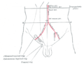
 Sigmoid colon and rectum, showing distribution of branches of inferior mesenteric artery and their anastomoses. (Inferior mesenteric artery labeled at center.) | |
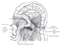
 Abdominal part of digestive tube and its attachment to the primitive or common mesentery. Human embryo of six weeks. (Inferior mesenteric artery labeled at bottom right.) | |
| Details | |
| Precursor | Vitelline arteries |
| Source | Abdominal aorta |
| Branches | Left colic artery, sigmoid branches, superior rectal artery |
| Vein | Inferior mesenteric vein |
| Supplies | Large Intestine |
| Identifiers | |
| Latin | arteria mesenterica inferior |
| MeSH | D017537 |
| TA98 | A12.2.12.069 |
| TA2 | 4291 |
| FMA | 14750 |
| Anatomical terminology | |
In human anatomy, the inferior mesenteric artery (IMA) is the third main branch of the abdominal aorta and arises at the level of L3, supplying the large intestine from the distal transverse colon to the upper part of the anal canal. The regions supplied by the IMA are the descending colon, the sigmoid colon, and part of the rectum. [1]


