Splenic artery

Last updated
Splenic artery
Gray1188.png
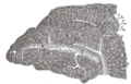
The visceral surface of the spleen.
Gray533.png
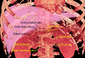
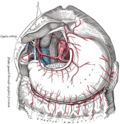
Branches of the celiac artery. (Lienal artery is an old term for splenic artery, and is visible at center. The spleen is at center right. The stomach has been flipped out to reveal the splenic artery, so the greater curvature is at the top in this diagram.)
Details
Source Celiac artery
Branches Pancreatic branches
pancreatica magna
left gastro-omental
short gastric
trabecular arteries
posterior gastric
Vein Splenic vein
Supplies Spleen
Identifiers
Latin arteria splenica, arteria lienalis
MeSH D013157
TA98 A12.2.12.040
TA2 4239
FMA 14773
Anatomical terminology

In human anatomy, the splenic artery or lienal artery, an older term, is the blood vessel that supplies oxygenated blood to the spleen. It branches from the celiac artery, and follows a course superior to the pancreas. It is known for its tortuous path to the spleen.

Contents

Structure

The splenic artery, the largest branch of the celiac trunk, gives off branches to the stomach and pancreas before reaching the spleen.

BranchDescription
branches to the pancreas multiple branches serving the pancreas including greater pancreatic artery and dorsal pancreatic artery.
short gastric upper part of greater curvature of the stomach and fundus of the stomach
left gastroepiploic middle of greater curvature of the stomach
posterior gastric posterior of stomach, gastric region superior to the splenic artery

Note that the branches of the splenic artery do not reach all the way to the lower part of the greater curvature of the stomach. Instead, that region is supplied by the right gastroepiploic artery, a branch of the gastroduodenal artery. The two gastroepiploic arteries anastomose with each other at that point.

Relations

The splenic artery passes between the layers of the lienorenal ligament. Along its course, it is accompanied by a similarly named vein, the splenic vein, which drains into the hepatic portal vein.

Clinical significance

Free fluid seen on ultrasound from a ruptured splenic artery aneurysm [1]

Splenic artery aneurysms are rare, but still the third most common abdominal aneurysm, after aneurysms of the abdominal aorta and iliac arteries. They may occur in pregnant women in the third trimester and rupture carries a maternal mortality of greater than 50% and a fetal mortality of 70 to 90%. Risk factors include smoking and hypertension. For the treatment of patients who represent a high surgical risk, percutaneous endovascular treatment may be considered.

Additional images

Related Research Articles

<span class="mw-page-title-main">Spleen</span> Organ recycling old red blood cells and also housing lymphocytes

The spleen is an organ found in almost all vertebrates. Similar in structure to a large lymph node, it acts primarily as a blood filter. The word spleen comes from Ancient Greek σπλήν (splḗn).

<span class="mw-page-title-main">Portal vein</span> Short thick vein formed by the union of the superior mesenteric vein and the splenic vein

The portal vein or hepatic portal vein (HPV) is a blood vessel that carries blood from the gastrointestinal tract, gallbladder, pancreas and spleen to the liver. This blood contains nutrients and toxins extracted from digested contents. Approximately 75% of total liver blood flow is through the portal vein, with the remainder coming from the hepatic artery proper. The blood leaves the liver to the heart in the hepatic veins.

<span class="mw-page-title-main">Abdominal aorta</span> Largest artery in the abdomen

In human anatomy, the abdominal aorta is the largest artery in the abdominal cavity. As part of the aorta, it is a direct continuation of the descending aorta.

<span class="mw-page-title-main">Renal artery</span> Vessel supplying blood to kidney

The renal arteries are paired arteries that supply the kidneys with blood. Each is directed across the crus of the diaphragm, so as to form nearly a right angle.

<span class="mw-page-title-main">Celiac artery</span> First major branch of the abdominal aorta

The celiacartery, also known as the celiac trunk or truncus coeliacus, is the first major branch of the abdominal aorta. It is about 1.25 cm in length. Branching from the aorta at thoracic vertebra 12 (T12) in humans, it is one of three anterior/ midline branches of the abdominal aorta.

<span class="mw-page-title-main">Superior mesenteric artery</span> Artery which supplies blood to the intestines and pancreas

In human anatomy, the superior mesenteric artery (SMA) is an artery which arises from the anterior surface of the abdominal aorta, just inferior to the origin of the celiac trunk, and supplies blood to the intestine from the lower part of the duodenum through two-thirds of the transverse colon, as well as the pancreas.

<span class="mw-page-title-main">Inferior mesenteric artery</span>

In human anatomy, the inferior mesenteric artery (IMA) is the third main branch of the abdominal aorta and arises at the level of L3, supplying the large intestine from the distal transverse colon to the upper part of the anal canal. The regions supplied by the IMA are the descending colon, the sigmoid colon, and part of the rectum.

<span class="mw-page-title-main">Common hepatic artery</span> Artery

The common hepatic artery is a short blood vessel that supplies oxygenated blood to the liver, pylorus of the stomach, duodenum, pancreas, and gallbladder.

<span class="mw-page-title-main">Superior mesenteric vein</span> Vein which drains blood from the small intestine

In human anatomy, the superior mesenteric vein (SMV) is a blood vessel that drains blood from the small intestine. Behind the neck of the pancreas, the superior mesenteric vein combines with the splenic vein to form the portal vein that carries blood to the liver. The superior mesenteric vein lies to the right of the similarly named artery, the superior mesenteric artery, which originates from the abdominal aorta.

<span class="mw-page-title-main">Splenic vein</span> Vein that drains blood from the spleen, stomach and pancreas

In human anatomy, the splenic vein is a blood vessel that drains blood from the spleen, the stomach fundus and part of the pancreas. It is part of the hepatic portal system.

<span class="mw-page-title-main">Left gastric artery</span> Artery

In human anatomy, the left gastric artery arises from the celiac artery and runs along the superior portion of the lesser curvature of the stomach before anastomosing with the right gastric artery. It also issues esophageal branches that supply lower esophagus and ascend through the esophageal hiatus to form anastomoses with the esophageal branches of thoracic part of aorta.

<span class="mw-page-title-main">Hepatic artery proper</span>

The hepatic artery proper is the artery that supplies the liver and gallbladder. It raises from the common hepatic artery, a branch of the celiac artery.

<span class="mw-page-title-main">Right gastroepiploic artery</span>

The right gastroepiploic artery is one of the two terminal branches of the gastroduodenal artery. It runs from right to left along the greater curvature of the stomach, between the layers of the greater omentum, anastomosing with the left gastroepiploic artery, a branch of the splenic artery.

<span class="mw-page-title-main">Left gastroepiploic artery</span>

The left gastroepiploic artery, the largest branch of the splenic artery, runs from left to right about a finger's breadth or more from the greater curvature of the stomach, between the layers of the greater omentum, and anastomoses with the right gastroepiploic.

<span class="mw-page-title-main">Superior pancreaticoduodenal artery</span> Artery

The superior pancreaticoduodenal artery is an artery that supplies blood to the duodenum and pancreas.

<span class="mw-page-title-main">Inferior pancreaticoduodenal artery</span> Branch of the superior mesenteric artery that supplies parts of the pancreas and the duodenum

The inferior pancreaticoduodenal artery is a branch of the superior mesenteric artery. It supplies the head of the pancreas, and the ascending and inferior parts of the duodenum. Rarely, it may have an aneurysm.

<span class="mw-page-title-main">Greater omentum</span> Fat sheath under abdominal wall

The greater omentum is a large apron-like fold of visceral peritoneum that hangs down from the stomach. It extends from the greater curvature of the stomach, passing in front of the small intestines and doubles back to ascend to the transverse colon before reaching to the posterior abdominal wall. The greater omentum is larger than the lesser omentum, which hangs down from the liver to the lesser curvature. The common anatomical term "epiploic" derives from "epiploon", from the Greek epipleein, meaning to float or sail on, since the greater omentum appears to float on the surface of the intestines. It is the first structure observed when the abdominal cavity is opened anteriorly.

<span class="mw-page-title-main">Hemosuccus pancreaticus</span> Medical condition

Hemosuccus pancreaticus is a rare cause of hemorrhage in the gastrointestinal tract. It is caused by a bleeding source in the pancreas, pancreatic duct, or structures adjacent to the pancreas, such as the splenic artery, that bleed into the pancreatic duct, which is connected with the bowel at the duodenum, the first part of the small intestine. Patients with hemosuccus may develop symptoms of gastrointestinal hemorrhage, such as blood in the stools, maroon stools, or melena, which is a dark, tarry stool caused by digestion of red blood cells. They may also develop abdominal pain. It is associated with pancreatitis, pancreatic cancer and aneurysms of the splenic artery. Hemosuccus may be identified with endoscopy (esophagogastroduodenoscopy), where fresh blood may be seen from the pancreatic duct. Alternatively, angiography may be used to inject the celiac axis to determine the blood vessel that is bleeding. This may also be used to treat hemosuccus, as embolization of the end vessel may terminate the hemorrhage. However, a distal pancreatectomy—surgery to remove of the tail of the pancreas—may be required to stop the hemorrhage.

<span class="mw-page-title-main">Celiac lymph nodes</span>

The celiac lymph nodes are associated with the branches of the celiac artery. Other lymph nodes in the abdomen are associated with the superior and inferior mesenteric arteries. The celiac lymph nodes are grouped into three sets: the gastric, hepatic and splenic lymph nodes. They receive lymph from the stomach, duodenum, pancreas, spleen, liver, and gall bladder.

The superior mesenteric vessels are composed of the superior mesenteric artery and the superior mesenteric vein.

References

  1. "UOTW #46 - Ultrasound of the Week". Ultrasound of the Week. 3 May 2015. Retrieved 27 May 2017.
  1. ^ Abbas MA, Stone WM, Fowl RJ, Gloviczki P, Oldenburg WA, Pairolero PC, Hallett JW, Bower TC, Panneton JM, Cherry KJ (2002)."Splenic arterya neurysms: two decades experience at Mayo clinic". Ann Vasc Surg.16(4): 442-449. PMID   12089631
  2. ^ Jamsheer NS, Malik M (2001). "Ruptured splenic artery aneurysm". Ann Saudi Med. 21(5-6): pp. 340–341. Accessed July 30, 2005.
  3. ^ Pauletto R, Mieres J, Incarbone A, García A, Santaera O (2013). "Exclusión de aneurisma esplénico gigante con técnica de catéteres mother-in-child en paciente de alto riesgo quirúrgico". Revista Argentina de Cardioangiología Vol. 4(1): pp. 62–65, Buenos Aires (Argentina): Colegio Argentino de Cardioangiología Intervencionista. ISSN 2313-9307, retrieved 11 September 2013.
  4. ^ Suzuki K, Prates JC, DiDio LJ (1978)."Incidence and surgical importance of the posterior gastricartery", Ann. Surg.187(2): 134-136.
  5. ^ Pauletto R, Mieres J, Incarbone A, García A, Santaera O (2013). "Exclusión de aneurisma esplénico gigante con técnica de catéteres mother-in-child en paciente de alto riesgo quirúrgico". Revista Argentina de Cardioangiología Vol. 4(1): pp. 62–65, Buenos Aires (Argentina): Colegio Argentino de Cardioangiología Intervencionista. ISSN 2313-9307, retrieved 11 September 2013.