
The anal canal is the part that connects the rectum to the anus, located below the level of the pelvic diaphragm. It is located within the anal triangle of the perineum, between the right and left ischioanal fossa. As the final functional segment of the bowel, it functions to regulate release of excrement by two muscular sphincter complexes. The anus is the aperture at the terminal portion of the anal canal.

In human anatomy, the inferior mesenteric artery (IMA) is the third main branch of the abdominal aorta and arises at the level of L3, supplying the large intestine from the distal transverse colon to the upper part of the anal canal. The regions supplied by the IMA are the descending colon, the sigmoid colon, and part of the rectum.

The facial artery is a branch of the external carotid artery that supplies structures of the superficial face.

The internal anal sphincter, IAS, or sphincter ani internus is a ring of smooth muscle that surrounds about 2.5–4.0 cm of the anal canal. It is about 5 mm thick, and is formed by an aggregation of the smooth (involuntary) circular muscle fibers of the rectum. It terminates distally about 6 mm from the anal orifice.

The internal iliac artery is the main artery of the pelvis.

In human anatomy, the inferior epigastric artery is an artery that arises from the external iliac artery. It is accompanied by the inferior epigastric vein; inferiorly, these two inferior epigastric vessels together travel within the lateral umbilical fold The inferior epigastric artery then traverses the arcuate line of rectus sheath to enter the rectus sheath, then anastomoses with the superior epigastric artery within the rectus sheath.

In human anatomy, the superior epigastric artery is a terminal branch of the internal thoracic artery that provides arterial supply to the abdominal wall, and upper rectus abdominis muscle. It enters the rectus sheath to descend upon the inner surface of the rectus abdominis muscle. It ends by anastomosing with the inferior epigastric artery.

The superior cerebellar artery (SCA) is an artery of the head. It arises near the end of the basilar artery. It is a branch of the basilar artery. It supplies parts of the cerebellum, the midbrain, and other nearby structures. It is the cause of trigeminal neuralgia in some patients.

The lingual artery arises from the external carotid artery between the superior thyroid artery and facial artery. It can be located easily in the tongue.

The uterine artery is an artery that supplies blood to the uterus in females.

The lateral sacral arteries is an artery in the pelvis that arises from the posterior division of the internal iliac artery. It later splits into two smaller branches, a superior and an inferior.

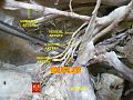
The inferior rectal artery is an artery that supplies blood to the lower third of the anal canal below the pectinate line.

The lateral circumflex femoral artery is an artery in the upper thigh. It is usually a branch of the profunda femoris artery, and produces three branches. It is mostly distributed to the muscles of the lateral thigh, supplying arterial blood to muscles of the knee extensor group.
The ovarian artery is an artery that supplies oxygenated blood to the ovary in females. It arises from the abdominal aorta below the renal artery. It can be found within the suspensory ligament of the ovary, anterior to the ovarian vein and ureter.

The lacrimal artery is an artery of the orbit. It is a branch of the ophthalmic artery. It accompanies the lacrimal nerve along the upper border of the lateral rectus muscle, travelling forward to reach the lacrimal gland. It supplies the lacrimal gland, two rectus muscles of the eye, the eyelids, and the conjunctiva.

The rectal venous plexus is the venous plexus surrounding the rectum. It consists of an internal and an external rectal plexus. It is drained by the superior, middle, and inferior rectal veins. It forms a portosystemic (portocaval) anastomosis. This allows rectally administered medications to bypass first pass metabolism.
The superior rectal artery is an artery that descends into the pelvis to supply blood to the rectum.
The inferior rectal nerves usually branch from the pudendal nerve but occasionally arises directly from the sacral plexus; they cross the ischiorectal fossa along with the inferior rectal artery and veins, toward the anal canal and the lower end of the rectum, and is distributed to the sphincter ani externus and to the integument (skin) around the anus.

The superior suprarenal artery is an artery in the abdomen. It is a branch of the inferior phrenic artery, itself a branch of the aorta. It supplies the adrenal gland.

The rectum is the final straight portion of the large intestine in humans and some other mammals, and the gut in others. The adult human rectum is about 12 centimetres (4.7 in) long, and begins at the rectosigmoid junction at the level of the third sacral vertebra or the sacral promontory depending upon what definition is used. Its diameter is similar to that of the sigmoid colon at its commencement, but it is dilated near its termination, forming the rectal ampulla. It terminates at the level of the anorectal ring or the dentate line, again depending upon which definition is used. In humans, the rectum is followed by the anal canal, which is about 4 centimetres (1.6 in) long, before the gastrointestinal tract terminates at the anal verge. The word rectum comes from the Latin rēctumintestīnum, meaning straight intestine.


