Common iliac artery

Last updated

Common iliac artery
Iliac artery bifurcation and aorta.PNG
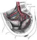
Front of abdomen, showing surface markings for arteries and inguinal canal.
Volume rendered CT scan of abdominal and pelvic blood vessels (smaller).gif
Volume rendered CT scan of abdominal and pelvic blood vessels.
Details
Source abdominal aorta
Branches external iliac
internal iliac
Vein common iliac veins
Identifiers
Latin arteria iliaca communis
TA98 A12.2.14.001
TA2 4301
FMA 14764
Anatomical terminology

The common iliac artery is a large artery of the abdomen paired on each side. It originates from the aortic bifurcation at the level of the 4th lumbar vertebra. It ends in front of the sacroiliac joint, one on either side, and each bifurcates into the external and internal iliac arteries.

Contents

Structure

The common iliac artery are about 4 cm long in adults and more than a centimeter in diameter. It begins as a branch of the aorta. [1] This is at the level of the 4th lumbar vertebra. [1] It runs inferolaterally, along the medial border of the psoas muscles. It bifurcates into the external iliac artery and the internal iliac artery at the pelvic brim, in front of the sacroiliac joints. [1]

The common iliac artery, and all of its branches, exist as paired structures (that is to say, there is one on the left side and one on the right).

The distribution of the common iliac artery is basically the pelvis and lower limb (as the femoral artery) on the corresponding side.

Relations

Both common iliac arteries are accompanied along their course by the two common iliac veins, which lie posteriorly and to the right. Their terminal bifurcation is crossed anteriorly by the ureters. [1] This is significant, as the bifurcation of the common iliac artery is the second point of ureteric constriction. [1]

Function

The common iliac artery supplies the leg and the pelvic region.[ citation needed ]

Clinical significance

Constriction

The common iliac artery may become narrowed. [2] This is most common at the aortic bifurcation. [2]

Dilatation

Dilatation of the common iliac artery can be graded into the following categories: [3]

NormalDiameter ≤ 12 mm
Ectasia Diameter 12 to 18 mm
Aneurysm Diameter ≥ 18 mm

Additional images

Related Research Articles

Aorta Largest artery in the body

The aorta is the main and largest artery in the human body, originating from the left ventricle of the heart and extending down to the abdomen, where it splits into two smaller arteries. The aorta distributes oxygenated blood to all parts of the body through the systemic circulation.

Inferior vena cava One of two veinous trunks bringing deoxygenated blood back to the heart

The inferior vena cava is a large vein that carries the deoxygenated blood from the lower and middle body into the right atrium of the heart. It is formed by the joining of the right and the left common iliac veins, usually at the level of the fifth lumbar vertebra.

Thoracic duct

In human anatomy, the thoracic duct is the larger of the two lymph ducts of the lymphatic system. It is also known as the left lymphatic duct, alimentary duct, chyliferous duct, and Van Hoorne's canal. The other duct is the right lymphatic duct. The thoracic duct carries chyle, a liquid containing both lymph and emulsified fats, rather than pure lymph. It also collects most of the lymph in the body other than from the right thorax, arm, head, and neck. The thoracic duct usually starts from the level of the twelfth thoracic vertebrae (T12) and extends to the root of the neck. It drains into the systemic (blood) circulation at the junction of the left subclavian and internal jugular veins, at the commencement of the brachiocephalic vein.

Celiac plexus Complex network of nerves located in the upper abdomen

The celiac plexus, also known as the solar plexus because of its radiating nerve fibers, is a complex network of nerves located in the abdomen, near where the celiac trunk, superior mesenteric artery, and renal arteries branch from the abdominal aorta. It is behind the stomach and the omental bursa, and in front of the crura of the diaphragm, on the level of the first lumbar vertebra.

Abdominal aorta

The abdominal aorta is the largest artery in the abdominal cavity. As part of the aorta, it is a direct continuation of the descending aorta.

Inferior mesenteric artery

In human anatomy, the inferior mesenteric artery, often abbreviated as IMA, is the third main branch of the abdominal aorta and arises at the level of L3, supplying the large intestine from the distal transverse colon to the upper part of the anal canal. The regions supplied by the IMA are the descending colon, the sigmoid colon, and part of the rectum.

External iliac artery Arteries of the pelvis

The external iliac arteries are two major arteries which bifurcate off the common iliac arteries anterior to the sacroiliac joint of the pelvis.

Abdominal external oblique muscle

The abdominal external oblique muscle is the largest and outermost of the three flat abdominal muscles of the lateral anterior abdomen.

Internal iliac artery

The internal iliac artery is the main artery of the pelvis.

Inferior epigastric artery Blood vessel

In human anatomy, inferior epigastric artery refers to the artery that arises from the external iliac artery. It anastomoses with the superior epigastric artery. Along its course, it is accompanied by a similarly named vein, the inferior epigastric vein. These epigastric vessels form the lateral border of Hesselbach's triangle, which outlines the area through which direct inguinal hernias protrude.

Iliohypogastric nerve

The iliohypogastric nerve is a nerve that originates from the lumbar plexus that supplies sensation to skin over the lateral gluteal and hypogastric regions and motor to the internal oblique muscles and transverse abdominal muscles.

Obturator nerve

The obturator nerve in human anatomy arises from the ventral divisions of the second, third, and fourth lumbar nerves in the lumbar plexus; the branch from the third is the largest, while that from the second is often very small.

Median sacral artery

The median sacral artery is a small artery that arises posterior to the abdominal aorta and superior to its bifurcation.

Lumbar arteries

The lumbar arteries are arteries located in the lower back or lumbar region. The lumbar arteries are in parallel with the intercostals.

Transversalis fascia Aponeurosis between the transverse abdominal muscle and the parietal peritoneum

The transversalis fascia is a thin aponeurotic membrane of the abdomen. It lies between the inner surface of the transverse abdominal muscle and the parietal peritoneum.

Superior hypogastric plexus

The superior hypogastric plexus is a plexus of nerves situated on the vertebral bodies anterior to the bifurcation of the abdominal aorta.

Superior rectal artery

The superior rectal artery is an artery that descends into the pelvis to supply blood to the rectum.

Hypogastric nerve

The hypogastric nerve is the nerve that transitions between the superior hypogastric plexus and the inferior hypogastric plexus. The hypogastric nerve enters the sympathetic chain at T12-L3.

Common iliac lymph nodes

The common iliac lymph nodes, four to six in number, are grouped behind and on the sides of the common iliac artery, one or two being placed below the bifurcation of the aorta, in front of the fifth lumbar vertebra.

Aortic bifurcation

The aortic bifurcation is the point at which the abdominal aorta bifurcates (forks) into the left and right common iliac arteries. The aortic bifurcation is usually seen at the level of L4, just above the junction of the left and right common iliac veins.

References

  1. 1 2 3 4 5 Jacob, S. (2008). "4 - Abdomen". Human Anatomy. Churchill Livingstone. pp. 71–123. doi:10.1016/B978-0-443-10373-5.50007-5. ISBN   978-0-443-10373-5.
  2. 1 2 Buckley, Brendan; Holden, Andrew; Merrilees, Stephen; Fernando, Rukshan (2020). "13 - Revascularization: Aortoiliac". Image-Guided Interventions - Expert Radiology (3rd ed.). Saunders. pp. 99–110. doi:10.1016/B978-0-323-61204-3.00013-0. ISBN   978-0-323-61204-3. S2CID   241869039.
  3. Melissa L Kirkwood. "Iliac artery aneurysm" . Retrieved February 23, 2018. Last updated: Mar 27, 2017.