Pelvic brim

Last updated
Pelvic brim
Pelvic inlet 03 animation.gif
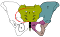
The edge of the pelvic inlet (shown in red) is the pelvic brim
Gray238.png
Diameters of superior aperture of lesser pelvis -- female. (Pelvic brim is not labeled, but is identifiable as the central opening at the top.)
Identifiers
FMA 224780
Anatomical terms of bone

The pelvic brim is the edge of the pelvic inlet. It is an approximately Mickey Mouse head-shaped line passing through the prominence of the sacrum, the arcuate and pectineal lines, and the upper margin of the pubic symphysis.

Contents

Structure

The pelvic brim is an approximately Mickey Mouse head-shaped line passing through the prominence of the sacrum, the arcuate and pectineal lines, and the upper margin of the pubic symphysis.

The pelvic brim is obtusely pointed in front, diverging on either side, and encroached upon behind by the projection forward of the promontory of the sacrum.

The oblique plane passing approximately through the pelvic brim divides the internal part of the pelvis (pelvic cavity) into the false or greater pelvis and the true or lesser pelvis. The false pelvis, which is above that plane, is sometimes considered to be a part of the abdominal cavity, rather than a part of the pelvic cavity. In this case, the pelvic cavity coincides with the true pelvis, which is below the above-mentioned plane.

The urinary bladder lies just above the anterior pelvic brim. [1] The sigmoid colon also passes close to the pelvic brim. [2]

Clinical significance

The pelvic brim may be a site of compression of structures that pass through the pelvic inlet. This can include the femoral nerves, which may occur during abdominal surgery. [3]

See also

Additional images

Related Research Articles

<span class="mw-page-title-main">Levator ani</span> Broad, thin muscle group, situated on either side of the pelvis

The levator ani is a broad, thin muscle group, situated on either side of the pelvis. It is formed from three muscle components: the pubococcygeus, the iliococcygeus, and the puborectalis.

<span class="mw-page-title-main">Sigmoid colon</span> Section of the large intestine closest to the rectum and anus

The sigmoid colon is the part of the large intestine that is closest to the rectum and anus. It forms a loop that averages about 35–40 centimetres (14–16 in) in length. The loop is typically shaped like a Greek letter sigma (ς) or Latin letter S. This part of the colon normally lies within the pelvis, but due to its freedom of movement it is liable to be displaced into the abdominal cavity.

<span class="mw-page-title-main">Pubic symphysis</span> Cartilaginous joint between the front of the left and right hip bones

The pubic symphysis is a secondary cartilaginous joint between the left and right superior rami of the pubis of the hip bones. It is in front of and below the urinary bladder. In males, the suspensory ligament of the penis attaches to the pubic symphysis. In females, the pubic symphysis is close to the clitoris. In most adults it can be moved roughly 2 mm and with 1 degree rotation. This increases for women at the time of childbirth.

<span class="mw-page-title-main">Pelvic floor</span> Anatomical structure

The pelvic floor or pelvic diaphragm is composed of muscle fibers of the levator ani, the coccygeus muscle, and associated connective tissue which span the area underneath the pelvis. The pelvic diaphragm is a muscular partition formed by the levatores ani and coccygei, with which may be included the parietal pelvic fascia on their upper and lower aspects. The pelvic floor separates the pelvic cavity above from the perineal region below. Both males and females have a pelvic floor. To accommodate the birth canal, a female's pelvic cavity is larger than a male's.

<span class="mw-page-title-main">Coccygeus muscle</span> Muscle of the lower back arising by its apex from the spine of the ischium

The coccygeus muscle or ischiococcygeus is a muscle of the pelvic floor, located posterior to levator ani and anterior to the sacrospinous ligament.

<span class="mw-page-title-main">Obturator internus muscle</span> One of six small hip muscles in the lateral rotator group

The internal obturator muscle or obturator internus muscle originates on the medial surface of the obturator membrane, the ischium near the membrane, and the rim of the pubis.

<span class="mw-page-title-main">Common iliac artery</span> Artery in the abdomen

The common iliac artery is a large artery of the abdomen paired on each side. It originates from the aortic bifurcation at the level of the 4th lumbar vertebra. It ends in front of the sacroiliac joint, one on either side, and each bifurcates into the external and internal iliac arteries.

<span class="mw-page-title-main">Anterior superior iliac spine</span> Bony projection of the iliac bone

The anterior superior iliac spine is a bony projection of the iliac bone, and an important landmark of surface anatomy. It refers to the anterior extremity of the iliac crest of the pelvis. It provides attachment for the inguinal ligament, and the sartorius muscle. The tensor fasciae latae muscle attaches to the lateral aspect of the superior anterior iliac spine, and also about 5 cm away at the iliac tubercle.

<span class="mw-page-title-main">Abdomen</span> Part of the body between the chest and pelvis

The abdomen is the part of the body between the thorax (chest) and pelvis, in humans and in other vertebrates. The abdomen is the front part of the abdominal segment of the torso. The area occupied by the abdomen is called the abdominal cavity. In arthropods it is the posterior tagma of the body; it follows the thorax or cephalothorax.

<span class="mw-page-title-main">Lumbar plexus</span>

The lumbar plexus is a web of nerves in the lumbar region of the body which forms part of the larger lumbosacral plexus. It is formed by the divisions of the first four lumbar nerves (L1-L4) and from contributions of the subcostal nerve (T12), which is the last thoracic nerve. Additionally, the ventral rami of the fourth lumbar nerve pass communicating branches, the lumbosacral trunk, to the sacral plexus. The nerves of the lumbar plexus pass in front of the hip joint and mainly support the anterior part of the thigh.

<span class="mw-page-title-main">Pubis (bone)</span> Most forward-facing of the three main regions making up the os coxa

In vertebrates, the pubic region is the most forward-facing of the three main regions making up the coxal bone. The left and right pubic regions are each made up of three sections, a superior ramus, inferior ramus, and a body.

<span class="mw-page-title-main">Pelvic cavity</span> Body cavity bounded by the bones of the pelvis

The pelvic cavity is a body cavity that is bounded by the bones of the pelvis. Its oblique roof is the pelvic inlet. Its lower boundary is the pelvic floor.

<span class="mw-page-title-main">Linea terminalis</span>

The linea terminalis or innominate line consists of the pubic crest, pectineal line, the arcuate line, the sacral ala, and the sacral promontory.

<span class="mw-page-title-main">Pelvic inlet</span> Plane representing the boundary between the pelvic and abdominal cavities

The pelvic inlet or superior aperture of the pelvis is a planar surface which defines the boundary between the pelvic cavity and the abdominal cavity. It is a major target of measurements of pelvimetry.

<span class="mw-page-title-main">Pubic tubercle</span> Bone

The pubic tubercle is a prominent tubercle on the superior ramus of the pubis bone of the pelvis.

<span class="mw-page-title-main">Perineal membrane</span> Anatomical term

The perineal membrane is an anatomical term for a fibrous membrane in the perineum. The term "inferior fascia of urogenital diaphragm", used in older texts, is considered equivalent to the perineal membrane.

<span class="mw-page-title-main">Pelvic outlet</span>

The lower circumference of the lesser pelvis is very irregular; the space enclosed by it is named the inferior aperture or pelvic outlet. It is an important component of pelvimetry.

<span class="mw-page-title-main">Outline of human anatomy</span> Overview of and topical guide to human anatomy

The following outline is provided as an overview of and topical guide to human anatomy:

<span class="mw-page-title-main">Hip bone</span> Bone of the pelvis

The hip bone is a large flat bone, constricted in the center and expanded above and below. In some vertebrates it is composed of three parts: the ilium, ischium, and the pubis.

<span class="mw-page-title-main">Pelvis</span> Lower part of the trunk of the human body between the abdomen and the thighs

The pelvis is the lower part of the trunk, between the abdomen and the thighs, together with its embedded skeleton.

References

PD-icon.svgThis article incorporates text in the public domain from page 238 of the 20th edition of Gray's Anatomy (1918)

  1. Wilson, David A., ed. (2012-01-01), "Ultrasound: Urinary Tract", Clinical Veterinary Advisor, Saint Louis: W.B. Saunders, pp. 836–838, doi:10.1016/b978-1-4160-9979-6.00269-5, ISBN   978-1-4160-9979-6 , retrieved 2021-01-30
  2. Jacob, S. (2008-01-01), Jacob, S. (ed.), "Chapter 4 - Abdomen", Human Anatomy, Churchill Livingstone, pp. 71–123, doi:10.1016/b978-0-443-10373-5.50007-5, ISBN   978-0-443-10373-5 , retrieved 2021-01-30
  3. Lalkhen, Abdul Ghaaliq (2015-01-01), Tubbs, R. Shane; Rizk, Elias; Shoja, Mohammadali M.; Loukas, Marios (eds.), "Chapter 37 - Perioperative Peripheral Nerve Injuries Associated with Surgical Positioning", Nerves and Nerve Injuries, San Diego: Academic Press, pp. 587–602, ISBN   978-0-12-802653-3 , retrieved 2021-01-30