The scapula, also known as the shoulder blade, is the bone that connects the humerus with the clavicle. Like their connected bones, the scapulae are paired, with each scapula on either side of the body being roughly a mirror image of the other. The name derives from the Classical Latin word for trowel or small shovel, which it was thought to resemble.

The sacrum, in human anatomy, is a large, triangular bone at the base of the spine that forms by the fusing of the sacral vertebrae (S1–S5) between ages 18 and 30.

The iliopsoas muscle refers to the joined psoas major and the iliacus muscles. The two muscles are separate in the abdomen, but usually merge in the thigh. They are usually given the common name iliopsoas. The iliopsoas muscle joins to the femur at the lesser trochanter. It acts as the strongest flexor of the hip.

The ilium is the uppermost and largest region of the coxal bone, and appears in most vertebrates including mammals and birds, but not bony fish. All reptiles have an ilium except snakes, although some snake species have a tiny bone which is considered to be an ilium.

The gluteal muscles, often called glutes, are a group of three muscles which make up the gluteal region commonly known as the buttocks: the gluteus maximus, gluteus medius and gluteus minimus. The three muscles originate from the ilium and sacrum and insert on the femur. The functions of the muscles include extension, abduction, external rotation, and internal rotation of the hip joint.

The ischium forms the lower and back region of the hip bone.
In vertebrates, the pubis or pubic bone forms the lower and anterior part of each side of the hip bone. The pubis is the most forward-facing of the three bones that make up the hip bone. The left and right pubic bones are each made up of three sections; a superior ramus, an inferior ramus, and a body.

The superior gluteal artery is the terminal branch of the posterior division of the internal iliac artery. It exits the pelvis through the greater sciatic foramen before splitting into a superficial branch and a deep branch.

The pelvic cavity is a body cavity that is bounded by the bones of the pelvis. Its oblique roof is the pelvic inlet. Its lower boundary is the pelvic floor.

The wing(ala)of ilium is the large expanded portion of the ilium, the bone which bounds the greater pelvis laterally. It presents for examination two surfaces—an external and an internal—a crest, and two borders—an anterior and a posterior.

The anterior inferior iliac spine (AIIS) is a bony eminence on the anterior border of the hip bone, or, more precisely, the wing of the ilium.
Medial to the anterior inferior iliac spine is a broad, shallow groove, over which the iliacus and psoas major muscles pass. This groove is bounded medially by an eminence, the iliopubic eminence, which marks the point of union of the ilium and pubis.

The iliac fossa is a large, smooth, concave surface on the internal surface of the ilium.

The iliolumbar ligament is a strong ligament which attaches medially to the transverse process of the 5th lumbar vertebra, and laterally to back of the inner lip of the iliac crest.

The lower circumference of the lesser pelvis is very irregular; the space enclosed by it is named the inferior aperture or pelvic outlet. It is an important component of pelvimetry.
The iliopectineal line is the border of the iliopubic eminence. It can be defined as a compound structure of the arcuate line and pectineal line. With the sacral promontory, it makes up the linea terminalis.

The hip bone is a large flat bone, constricted in the center and expanded above and below. In some vertebrates it is composed of three parts: the ilium, ischium, and the pubis.

The pelvis is the lower part of an anatomical trunk, between the abdomen and the thighs, together with its embedded skeleton.
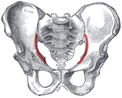
The gluteal lines are three curved lines outlined from three bony ridges on the exterior surface of the ilium in the gluteal region. They are the anterior gluteal line; the inferior gluteal line, and the posterior gluteal line.
X-rays of hip dysplasia are one of the two main methods of medical imaging to diagnose hip dysplasia, the other one being medical ultrasonography. Ultrasound imaging yields better results defining the anatomy until the cartilage is ossified. When the infant is around 3 months old a clear roentgenographic image can be achieved. Unfortunately the time the joint gives a good x-ray image is also the point at which nonsurgical treatment methods cease to give good results.