
The pudendal nerve is the main nerve of the perineum. It is a mixed nerve and also conveys sympathetic autonomic fibers. It carries sensation from the external genitalia of both sexes and the skin around the anus and perineum, as well as the motor supply to various pelvic muscles, including the male or female external urethral sphincter and the external anal sphincter.
Pudendal nerve entrapment (PNE), also known as Alcock canal syndrome, is an uncommon source of chronic pain in which the pudendal nerve is entrapped or compressed in Alcock's canal. There are several different types of PNE based on the site of entrapment anatomically. Pain is positional and is worsened by sitting. Other symptoms include genital numbness, fecal incontinence and urinary incontinence.

The internal pudendal artery is one of the three pudendal arteries. It branches off the internal iliac artery, and provides blood to the external genitalia.

The piriformis muscle is a flat, pyramidally-shaped muscle in the gluteal region of the lower limbs. It is one of the six muscles in the lateral rotator group.

The internal obturator muscle or obturator internus muscle originates on the medial surface of the obturator membrane, the ischium near the membrane, and the rim of the pubis.

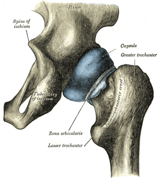
The ischium forms the lower and back region of the hip bone.
In vertebrates, the pubis or pubic bone forms the lower and anterior part of each side of the hip bone. The pubis is the most forward-facing of the three bones that make up the hip bone. The left and right pubic bones are each made up of three sections, a superior ramus, inferior ramus, and a body.

The sacrotuberous ligament is situated at the lower and back part of the pelvis. It is flat, and triangular in form; narrower in the middle than at the ends.
The sacrospinous ligament is a thin, triangular ligament in the human pelvis. The base of the ligament is attached to the outer edge of the sacrum and coccyx, and the tip of the ligament attaches to the spine of the ischium, a bony protuberance on the human pelvis. Its fibres are intermingled with the sacrotuberous ligament.

The inferior gluteal artery is a terminal branch of the anterior trunk of the internal iliac artery. It exits the pelvis through the greater sciatic foramen. It is distributed chiefly to the buttock and the back of the thigh.
The greater sciatic foramen is an opening in the posterior human pelvis. It is formed by the sacrotuberous and sacrospinous ligaments. The piriformis muscle passes through the foramen and occupies most of its volume. The greater sciatic foramen is wider in women than in men.
The ischial tuberosity, also known colloquially as the sit bones or sitz bones, or as a pair the sitting bones, is a large swelling posteriorly on the superior ramus of the ischium. It marks the lateral boundary of the pelvic outlet.

The nerve to obturator internus is a mixed nerve providing motor innervation to the obturator internus muscle and gemellus superior muscle, and sensory innervation to the hip joint. It is a branch of the sacral plexus. It is one of the group of deep gluteal nerves.

The greater sciatic notch is a notch in the ilium, one of the bones that make up the human pelvis. It lies between the posterior inferior iliac spine (above), and the ischial spine (below). The sacrospinous ligament changes this notch into an opening, the greater sciatic foramen.

Below the ischial spine is a small notch, the lesser sciatic notch; it is smooth, coated in the recent state with cartilage, the surface of which presents two or three ridges corresponding to the subdivisions of the tendon of the obturator internus, which winds over it.

The anal triangle is the posterior part of the perineum. It contains the anal canal.

The lower circumference of the lesser pelvis is very irregular; the space enclosed by it is named the inferior aperture or pelvic outlet. It is an important component of pelvimetry.

The following outline is provided as an overview of and topical guide to human anatomy:

The hip bone is a large flat bone, constricted in the center and expanded above and below. In some vertebrates it is composed of three parts: the ilium, ischium, and the pubis.

The pelvis is the lower part of the trunk, between the abdomen and the thighs, together with its embedded skeleton.

