Related Research Articles

The gluteus maximus is the main extensor muscle of the hip. It is the largest and outermost of the three gluteal muscles and makes up a large part of the shape and appearance of each side of the hips. It is the single largest muscle in the human body. Its thick fleshy mass, in a quadrilateral shape, forms the prominence of the buttocks. The other gluteal muscles are the medius and minimus, and sometimes informally these are collectively referred to as the glutes.

The gluteus medius, one of the three gluteal muscles, is a broad, thick, radiating muscle. It is situated on the outer surface of the pelvis.

The pectineus muscle is a flat, quadrangular muscle, situated at the anterior (front) part of the upper and medial (inner) aspect of the thigh. The pectineus muscle is the most anterior adductor of the hip. The muscle does adduct and internally rotate the thigh but its primary function is hip flexion.

The anal canal is the part that connects the rectum to the anus, located below the level of the pelvic diaphragm. It is located within the anal triangle of the perineum, between the right and left ischioanal fossa. As the final functional segment of the bowel, it functions to regulate release of excrement by two muscular sphincter complexes. The anus is the aperture at the terminal portion of the anal canal.

The iliopsoas muscle refers to the joined psoas major and the iliacus muscles. The two muscles are separate in the abdomen, but usually merge in the thigh. They are usually given the common name iliopsoas. The iliopsoas muscle joins to the femur at the lesser trochanter. It acts as the strongest flexor of the hip.

The ilium is the uppermost and largest part of the hip bone, and appears in most vertebrates including mammals and birds, but not bony fish. All reptiles have an ilium except snakes, although some snake species have a tiny bone which is considered to be an ilium.

In vertebrates, the pubic region is the most forward-facing of the three main regions making up the coxal bone. The left and right pubic regions are each made up of three sections, a superior ramus, inferior ramus, and a body.

The suspensory ligament of the ovary, also infundibulopelvic ligament, is a fold of peritoneum that extends out from the ovary to the wall of the pelvis.

The pelvic cavity is a body cavity that is bounded by the bones of the pelvis. Its oblique roof is the pelvic inlet. Its lower boundary is the pelvic floor.

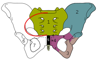
The linea terminalis or innominate line consists of the pubic crest, pectineal line, the arcuate line, the sacral ala, and the sacral promontory.

The arcuate line of the ilium is a smooth rounded border on the internal surface of the ilium. It is immediately inferior to the iliac fossa and Iliacus muscle.

At the level of a line extending from the lower part of the pubic symphysis to the spine of the ischium is a thickened whitish band in this upper layer of the diaphragmatic part of the pelvic fascia. It is termed the tendinous arch or white line of the pelvic fascia, and marks the line of attachment of the special fascia which is associated with the pelvic viscera. It joins the fascia of the pubocervical fascia that covers the anterior wall of the vagina. If this fascia falls, the ipsilateral side of the vagina falls, carrying with it the bladder and the urethra, and thus contributing to urinary incontinence.

Medial to the anterior inferior iliac spine is a broad, shallow groove, over which the iliacus and psoas major muscles pass. This groove is bounded medially by an eminence, the iliopubic eminence, which marks the point of union of the ilium and pubis.
Pelvis justo major is a rare condition of the adult female pelvis where the pelvis flairs above the Iliopectineal line. It is 1.5 or more times larger than an average pelvis in every direction and is at least 42 cm (16.5 inches) biiliac width. Even though this condition is classified as a congenital abnormality, it is not a medical disease or abnormality of the pelvis.

The pectineal line of the pubis is a ridge on the superior ramus of the pubic bone. It forms part of the pelvic brim.

The pelvic fasciae are the fascia of the pelvis and can be divided into:
The vascular lacuna is the compartment beneath the inguinal ligament which allows for passage of the femoral vessels, lymph vessels and lymph nodes. Its boundaries are the iliopectineal arch, the inguinal ligament, the lacunar ligament, and the superior border of the pubis. The structures found in the vascular lacuna, from medial to lateral, are:
The Iliopectineal arch is a thickened band of fused iliac fascia and psoas fascia passing from the posterior aspect of the inguinal ligament anteriorly across the front of the femoral nerve to attach to the iliopubic eminence of the hip bone posteriorly. The iliopectineal arch thus forms a septum which subdivides the space deep to the inguinal ligament into a lateral muscular lacuna and a medial vascular lacuna. When a psoas minor muscle is present, its tendon of insertion blends with the iliopectineal arch

The pelvis is the lower part of the trunk, between the abdomen and the thighs, together with its embedded skeleton.
X-rays of hip dysplasia are one of the two main methods of medical imaging to diagnose hip dysplasia, the other one being medical ultrasonography. Ultrasound imaging yields better results defining the anatomy until the cartilage is ossified. When the infant is around 3 months old a clear roentgenographic image can be achieved. Unfortunately the time the joint gives a good x-ray image is also the point at which nonsurgical treatment methods cease to give good results.
References
- ↑ Stedman's Medical dictionary 1914 3rd ed. Lippincott Williams & Wilkins. 1914. pp. 692–.
- ↑ Kirschner, Celeste G. (2005). Netter's Atlas Of Human Anatomy For CPT Coding. Chicago: American medical association. p. 274. ISBN 1-57947-669-4.