
The coccyx, commonly referred to as the tailbone, is the final segment of the vertebral column in all apes, and analogous structures in certain other mammals such as horses. In tailless primates since Nacholapithecus, the coccyx is the remnant of a vestigial tail. In animals with bony tails, it is known as tailhead or dock, in bird anatomy as tailfan. It comprises three to five separate or fused coccygeal vertebrae below the sacrum, attached to the sacrum by a fibrocartilaginous joint, the sacrococcygeal symphysis, which permits limited movement between the sacrum and the coccyx.

The sacrum, in human anatomy, is a large, triangular bone at the base of the spine that forms by the fusing of the sacral vertebrae (S1–S5) between ages 18 and 30.

The gluteus maximus is the main extensor muscle of the hip in humans. It is the largest and outermost of the three gluteal muscles and makes up a large part of the shape and appearance of each side of the hips. It is the single largest muscle in the human body. Its thick fleshy mass, in a quadrilateral shape, forms the prominence of the buttocks. The other gluteal muscles are the medius and minimus, and sometimes informally these are collectively referred to as the glutes.

The piriformis muscle is a flat, pyramidally-shaped muscle in the gluteal region of the lower limbs. It is one of the six muscles in the lateral rotator group.
The biceps femoris is a muscle of the thigh located to the posterior, or back. As its name implies, it consists of two heads; the long head is considered part of the hamstring muscle group, while the short head is sometimes excluded from this characterization, as it only causes knee flexion and is activated by a separate nerve.

The adductor magnus is a large triangular muscle, situated on the medial side of the thigh.
The semimembranosus muscle is the most medial of the three hamstring muscles in the thigh. It is so named because it has a flat tendon of origin. It lies posteromedially in the thigh, deep to the semitendinosus muscle. It extends the hip joint and flexes the knee joint.

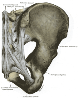
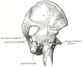
The sacrospinous ligament is a thin, triangular ligament in the human pelvis. The base of the ligament is attached to the outer edge of the sacrum and coccyx, and the tip of the ligament attaches to the spine of the ischium, a bony protuberance on the human pelvis. Its fibres are intermingled with the sacrotuberous ligament.

The fascia lata is the deep fascia of the thigh. It encloses the thigh muscles and forms the outer limit of the fascial compartments of thigh, which are internally separated by the medial intermuscular septum and the lateral intermuscular septum. The fascia lata is thickened at its lateral side where it forms the iliotibial tract, a structure that runs to the tibia and serves as a site of muscle attachment.

The inferior gluteal artery is a terminal branch of the anterior trunk of the internal iliac artery. It exits the pelvis through the greater sciatic foramen. It is distributed chiefly to the buttock and the back of the thigh.

The inferior rectal artery is an artery that supplies blood to the lower third of the anal canal below the pectinate line.

The internal pudendal veins are a set of veins in the pelvis. They are the venae comitantes of the internal pudendal artery. Internal pudendal veins are enclosed by pudendal canal, with internal pudendal artery and pudendal nerve.
The ischial tuberosity, also known colloquially as the sit bones or sitz bones, or as a pair the sitting bones, is a large posterior bony protuberance on the superior ramus of the ischium. It marks the lateral boundary of the pelvic outlet.

The greater sciatic notch is a notch in the ilium, one of the bones that make up the human pelvis. It lies between the posterior inferior iliac spine (above), and the ischial spine (below). The sacrospinous ligament changes this notch into an opening, the greater sciatic foramen.

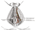
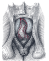
The urogenital triangle is the anterior part of the perineum. In female mammals, it contains the vulva, while in male mammals, it contains the penis and scrotum.

The anal triangle is the posterior part of the perineum. It contains the anus in mammals.

The following outline is provided as an overview of and topical guide to human anatomy:

The hip bone is a large flat bone, constricted in the center and expanded above and below. In some vertebrates it is composed of three parts: the ilium, ischium, and the pubis.

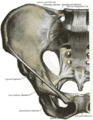
The pelvis is the lower part of an anatomical trunk, between the abdomen and the thighs, together with its embedded skeleton.

The gemelli muscles are the inferior gemellus muscle and the superior gemellus muscle, two small accessory fasciculi to the tendon of the internal obturator muscle. The gemelli muscles belong to the lateral rotator group of six muscles of the hip that rotate the femur in the hip joint.
