
The human leg is the entire lower limb of the human body, including the foot, thigh or sometimes even the hip or buttock region. The major bones of the leg are the femur, tibia, and adjacent fibula. The thigh is between the hip and knee, while the calf (rear) and shin (front) are between the knee and foot.
Articles related to anatomy include:

The piriformis muscle is a flat, pyramidally-shaped muscle in the gluteal region of the lower limbs. It is one of the six muscles in the lateral rotator group.

The internal obturator muscle or obturator internus muscle originates on the medial surface of the obturator membrane, the ischium near the membrane, and the rim of the pubis.
The external obturator muscle, obturator externus muscle is a flat, triangular muscle, which covers the outer surface of the anterior wall of the pelvis.
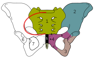
The obturator foramen is the large, bilaterally paired opening of the bony pelvis. It is formed by the pubis and ischium. It is mostly closed by the obturator membrane except for a small opening, the obturator canal, through which the obturator nerve and vessels pass.

The ischium forms the lower and back region of the hip bone.
In vertebrates, the pubis or pubic bone forms the lower and anterior part of each side of the hip bone. The pubis is the most forward-facing of the three bones that make up the hip bone. The left and right pubic bones are each made up of three sections, a superior ramus, inferior ramus, and a body.

The obturator artery is a branch of the internal iliac artery that passes antero-inferiorly on the lateral wall of the pelvis, to the upper part of the obturator foramen, and, escaping from the pelvic cavity through the obturator canal, it divides into an anterior branch and a posterior branch.

The basilar part of the occipital bone extends forward and upward from the foramen magnum, and presents in front an area more or less quadrilateral in outline.

The squamous part of occipital bone is situated above and behind the foramen magnum, and is curved from above downward and from side to side.

The acetabular notch is a deep notch in the inferior portion of the rim of the acetabulum. It is bridged by the transverse acetabular ligament, converting it into a foramen. It is continuous with space of the acetabular fossa. The lunate surface of acetabulum is discontinued opposite the notch.

The obturator fascia, or fascia of the internal obturator muscle, covers the pelvic surface of that muscle and is attached around the margin of its origin.

The nerve to obturator internus is a mixed nerve providing motor innervation to the obturator internus muscle and gemellus superior muscle, and sensory innervation to the hip joint. It is a branch of the sacral plexus. It is one of the group of deep gluteal nerves.

The tectorial membrane of atlanto-axial joint is a tough membrane/broad, strong band representing the superior-ward prolongation of the posterior longitudinal ligament.

The anterior border of the superior pubic ramus presents a sharp margin, the obturator crest, which forms part of the circumference of the obturator foramen superiorly and affords attachment to the obturator membrane.
The ischiopubic ramus is a compound structure consisting of the following two structures:
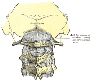
The posterior atlantooccipital membrane is a broad but thin membrane extending between the to the posterior margin of the foramen magnum above, and posterior arch of atlas below. It forms the floor of the suboccipital triangle.

The following outline is provided as an overview of and topical guide to human anatomy:

The hip bone is a large flat bone, constricted in the center and expanded above and below. In some vertebrates it is composed of three parts: the ilium, ischium, and the pubis.
