Ischioanal fossa

Last updated
Ischioanal fossa
Gray405.png
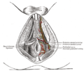
The perineum. The integument and superficial layer of superficial fascia reflected. (Ischiorectal fossa labeled at bottom left.)
Gray1077.png
The posterior aspect of the rectum exposed by removing the lower part of the sacrum and the coccyx (ischiorectal fossa labeled at bottom right)
Details
Identifiers
Latin fossa ischioanalis
TA98 A09.5.04.001
TA2 2446
FMA 22059
Anatomical terminology

The ischioanal fossa (formerly called ischiorectal fossa) is the fat-filled wedge-shaped space located lateral to the anal canal and inferior to the pelvic diaphragm. It is somewhat prismatic in shape, with its base directed to the surface of the perineum and its apex at the line of meeting of the obturator and anal fasciae.

Contents

Boundaries

It has the following boundaries: [1]

ANTERIOR
* fascia of Colles covering the transversus perinei superficialis
* inferior fascia of the urogenital diaphragm (perineal membrane)
LATERAL
* tuberosity of the ischium
* Obturator internus muscle
* obturator fascia
SUPERIOR:
* Levator ani

INFERIOR:
* skin

MEDIAL:
* Levator ani
* Sphincter ani externus muscle
* anal fascia
POSTERIOR
* Gluteus maximus
* sacrotuberous ligament

Contents

The contents include:

See also

Related Research Articles

<span class="mw-page-title-main">Pudendal nerve</span> Main nerve of the perineum

The pudendal nerve is the main nerve of the perineum. It is a mixed nerve and also conveys sympathetic autonomic fibers. It carries sensation from the external genitalia of both sexes and the skin around the anus and perineum, as well as the motor supply to various pelvic muscles, including the male or female external urethral sphincter and the external anal sphincter.

<span class="mw-page-title-main">Levator ani</span> Broad, thin muscle group, situated on either side of the pelvis

The levator ani is a broad, thin muscle group, situated on either side of the pelvis. It is formed from three muscle components: the pubococcygeus, the iliococcygeus, and the puborectalis.

<span class="mw-page-title-main">Ischiocavernosus muscle</span> Sheet of fibrous tissue between the thigh and the front of the perineum

The ischiocavernosus muscle is a muscle just below the surface of the perineum, present in both men and women.

<span class="mw-page-title-main">Bulbospongiosus muscle</span> Superficial muscle of the perineum

The bulbospongiosus muscles are a subgroup of the superficial muscles of the perineum. They have a slightly different origin, insertion and function in males and females. In males, these muscles cover the bulb of the penis, while in females, they cover the vestibular bulbs.

<span class="mw-page-title-main">Internal pudendal artery</span> Blood vessel supplying blood to the external genitalia

The internal pudendal artery is one of the three pudendal arteries. It branches off the internal iliac artery, and provides blood to the external genitalia.

<span class="mw-page-title-main">Anal canal</span> Functional segment of the large intestine

The anal canal is the part that connects the rectum to the anus, located below the level of the pelvic diaphragm. It is located within the anal triangle of the perineum, between the right and left ischioanal fossa. As the final functional segment of the bowel, it functions to regulate release of excrement by two muscular sphincter complexes. The anus is the aperture at the terminal portion of the anal canal.

<span class="mw-page-title-main">External iliac artery</span> Arteries of the pelvis

The external iliac arteries are two major arteries which bifurcate off the common iliac arteries anterior to the sacroiliac joint of the pelvis.

<span class="mw-page-title-main">Internal iliac artery</span> Main artery of the pelvis

The internal iliac artery is the main artery of the pelvis.

<span class="mw-page-title-main">Inferior epigastric artery</span> Blood vessel

In human anatomy, the inferior epigastric artery is an artery that arises from the external iliac artery. It is accompanied by the inferior epigastric vein; inferiorly, these two inferior epigastric vessels together travel within the lateral umbilical fold The inferior epigastric artery then traverses the arcuate line of rectus sheath to enter the rectus sheath, then anastomoses with the superior epigastric artery within the rectus sheath.

<span class="mw-page-title-main">Sacrotuberous ligament</span>

The sacrotuberous ligament is situated at the lower and back part of the pelvis. It is flat, and triangular in form; narrower in the middle than at the ends.

<span class="mw-page-title-main">Inferior rectal artery</span>

The inferior rectal artery is an artery that supplies blood to the lower third of the anal canal below the pectinate line.

<span class="mw-page-title-main">Perineal nerve</span> Nerve of the perineum

The perineal nerve is a nerve of the pelvis. It arises from the pudendal nerve in the pudendal canal. It gives superficial branches to the skin, and a deep branch to muscles. It supplies the skin and muscles of the perineum. Its latency is tested with electrodes.

<span class="mw-page-title-main">Internal pudendal veins</span> Set of veins in the pelvis enclosed by pudendal canal

The internal pudendal veins are a set of veins in the pelvis. They are the venae comitantes of the internal pudendal artery. Internal pudendal veins are enclosed by pudendal canal, with internal pudendal artery and pudendal nerve.

<span class="mw-page-title-main">Pudendal canal</span>

The pudendal canal is an anatomical structure formed by the obturator fascia lining the lateral wall of the ischioanal fossa. The internal pudendal artery and veins, and pudendal nerve pass through the pudendal canal, and the perineal nerve arises within it.

<span class="mw-page-title-main">Pelvic cavity</span> Body cavity bounded by the pelvic bones

The pelvic cavity is a body cavity that is bounded by the bones of the pelvis. Its oblique roof is the pelvic inlet. Its lower boundary is the pelvic floor.

<span class="mw-page-title-main">Superior rectal artery</span>

The superior rectal artery is an artery that descends into the pelvis to supply blood to the rectum.

<span class="mw-page-title-main">Inferior anal nerves</span>

The inferior rectal nerves usually branch from the pudendal nerve but occasionally arises directly from the sacral plexus; they cross the ischiorectal fossa along with the inferior rectal artery and veins, toward the anal canal and the lower end of the rectum, and is distributed to the sphincter ani externus and to the integument (skin) around the anus.

<span class="mw-page-title-main">Medial umbilical ligament</span>

The medial umbilical ligament, cord of umbilical artery, or obliterated umbilical artery is a paired structure found in human anatomy. It is on the deep surface of the anterior abdominal wall, and is covered by the medial umbilical folds. It is different from the median umbilical ligament, a structure that represents the remnant of the embryonic urachus.

<span class="mw-page-title-main">Deep perineal pouch</span> Anatomic space enclosed partly by the perineum

The deep perineal pouch is the anatomic space enclosed in part by the perineum and located superior to the perineal membrane.

<span class="mw-page-title-main">Anal triangle</span> Posterior part of the perineum

The anal triangle is the posterior part of the perineum. It contains the anal canal.

References

PD-icon.svgThis article incorporates text in the public domain from page 425 of the 20th edition of Gray's Anatomy (1918)

  1. analtrianglesection - Ischiorectal fossa is colored yellow

Additional images