Anal fascia

Last updated
Anal fascia

Gray402.png

Coronal section of pelvis, showing arrangement of fasciae. Viewed from behind.
Identifiers

Anatomical terminology

The anal fascia is the inferior layer of the diaphragmatic part of the pelvic fascia, which covers both surfaces of the levatores ani. It is attached above to the obturator fascia along the line of origin of the levator ani, while below it is continuous with the superior fascia of the urogenital diaphragm, and with the fascia on the sphincter ani internus.

Pelvic fascia

The pelvic fasciae are the fascia of the pelvis and can be divided into:

Levator ani

The levator ani is a broad, thin muscle, situated on either side of the pelvis. It is formed from three muscle components: the pubococcygeus, the iliococcygeus, and the puborectalis.

Obturator fascia

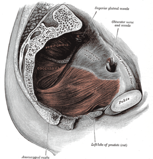
The obturator fascia, or fascia of the internal obturator muscle, covers the pelvic surface of that muscle and is attached around the margin of its origin.

The layer covering the upper surface of the pelvic diaphragm follows, above, the line of origin of the levator ani and is therefore somewhat variable.

In front it is attached to the back of the pubic symphysis about 2 cm. above its lower border.

Pubic symphysis

The pubic symphysis a cartilaginous joint that sits between and joins left and right the superior rami of the pubic bones. It is located in front of and below the urinary bladder. In males, the suspensory ligament of the penis attaches to the pubic symphysis. In females, the pubic symphysis is intimately close to the clitoris. In normal adults it can be moved roughly 2 mm and with 1 degree rotation. This increases for women at the time of childbirth.

It can then be traced laterally across the back of the superior ramus of the pubic bone for a distance of about 1.25 cm, when it reaches the obturator fascia.

Superior pubic ramus

The superior pubic ramus is a part of the pubic bone which forms a portion of the obturator foramen. The obturator foramen, along with the ilium and other fused bones, forms part of either side of the pelvis.

Pubis (bone) the ventral and anterior of the three principal bones composing either half of the pelvis

In vertebrates, the pubic bone is the ventral and anterior of the three principal bones composing either half of the pelvis.

It is attached to this fascia along a line which pursues a somewhat irregular course to the spine of the ischium.

Ischial spine

From the posterior border of the body of the Ischium there extends backward a thin and pointed triangular eminence, the ischial spine, more or less elongated in different subjects.

Ischium lower and back part of the hip bone

The ischium forms the lower and back part of the hip bone.

The irregularity of this line is because the origin of the levator ani, which in lower forms is from the pelvic brim, is in man lower down, on the obturator fascia.

Tendinous fibers of origin of the muscle are therefore often found extending up toward, and in some cases reaching, the pelvic brim, and on these the fascia is carried.

See also

Related Research Articles

Perineum region of the body including the perineal body and surrounding structures

The perineum is the space between the anus and scrotum in the male and between the anus and the vulva in the female. The perineum is the region of the body between the pubic symphysis and the coccyx, including the perineal body and surrounding structures. There is some variability in how the boundaries are defined. The perianal area is a subset of the perineum.

Pelvic floor anatomical structure

The pelvic floor or pelvic diaphragm is composed of muscle fibers of the levator ani, the coccygeus muscle, and associated connective tissue which span the area underneath the pelvis. The pelvic diaphragm is a muscular partition formed by the levatores ani and coccygei, with which may be included the parietal pelvic fascia on their upper and lower aspects. The pelvic floor separates the pelvic cavity above from the perineal region below. Because, to accommodate the birth canal, a female's pelvic cavity is larger than a male's, the pelvic floor tends to be considered a part of female anatomy, but males have an equivalent pelvic floor.

Internal obturator muscle

The internal obturator muscle or obturator internus muscle originates on the medial surface of the obturator membrane, the ischium near the membrane, and the rim of the pubis.

External sphincter muscle of male urethra muscle of the human male urethra

The external sphincter muscle of urethra, also sphincter urethrae membranaceae, sphincter urethrae externus, surrounds the whole length of the membranous urethra, and is enclosed in the fascia of the urogenital diaphragm.

External obturator muscle

The external obturator muscle, obturator externus muscle is a flat, triangular muscle, which covers the outer surface of the anterior wall of the pelvis.

Obturator artery

The obturator artery is a branch of the internal iliac artery that passes antero-inferiorly on the lateral wall of the pelvis, to the upper part of the obturator foramen, and, escaping from the pelvic cavity through the obturator canal, it divides into both an anterior and a posterior branch.

Pelvic cavity body cavity bounded by the bones of the pelvis

The pelvic cavity is a body cavity that is bounded by the bones of the pelvis. Its oblique roof is the pelvic inlet. Its lower boundary is the pelvic floor.

Perineal membrane

The perineal membrane is an anatomical term for a fibrous membrane in the perineum. The term "inferior fascia of urogenital diaphragm", used in older texts, is considered equivalent to the perineal membrane.

Inferior pubic ramus

The inferior pubic ramus is a part of the pelvis and is thin and flat. It passes laterally and downward from the medial end of the superior ramus; it becomes narrower as it descends and joins with the inferior ramus of the ischium below the obturator foramen.

Superior fascia of the urogenital diaphragm

The superior fascia of the urogenital diaphragm is continuous with the obturator fascia and stretches across the pubic arch.

Outline of human anatomy scientific study of the morphology of the human body

The following outline is provided as an overview of and topical guide to human anatomy:

Hip bone bone of the pelvis

The hip bone is a large irregular bone, constricted in the center and expanded above and below. In some vertebrates it is composed of three parts: the ilium, ischium, and the pubis.

Pelvis lower part of the trunk of the human body between the abdomen and the thighs (sometimes also called pelvic region of the trunk

The pelvis is either the lower part of the trunk of the human body between the abdomen and the thighs or the skeleton embedded in it.

Vaginal support structures

The vaginal support structures are those muscles, bones, ligaments, tendons, membranes and fascia, of the pelvic floor that maintain the position of the vagina within the pelvic cavity and allow the normal functioning of the vagina and other reproductive structures in the female. Defects or injuries to these support structures in the pelvic floor leads to pelvic organ prolapse. Anatomical and congenital variations of vaginal support structures can predispose a woman to further dysfunction and prolapse later in life. The urethra is part of the anterior wall of the vagina and damage to the support structures there can lead to incontinence and urinary retention.

References

This article incorporates text in the public domain from page 421 of the 20th edition of Gray's Anatomy (1918)

The public domain consists of all the creative works to which no exclusive intellectual property rights apply. Those rights may have expired, been forfeited, expressly waived, or may be inapplicable.

<i>Grays Anatomy</i> English-language textbook of human anatomy

Gray's Anatomy is an English language textbook of human anatomy originally written by Henry Gray and illustrated by Henry Vandyke Carter. Earlier editions were called Anatomy: Descriptive and Surgical and Gray's Anatomy: Descriptive and Applied, but the book's name is commonly shortened to, and later editions are titled, Gray's Anatomy. The book is widely regarded as an extremely influential work on the subject, and has continued to be revised and republished from its initial publication in 1858 to the present day. The latest edition of the book, the 41st, was published in September 2015.