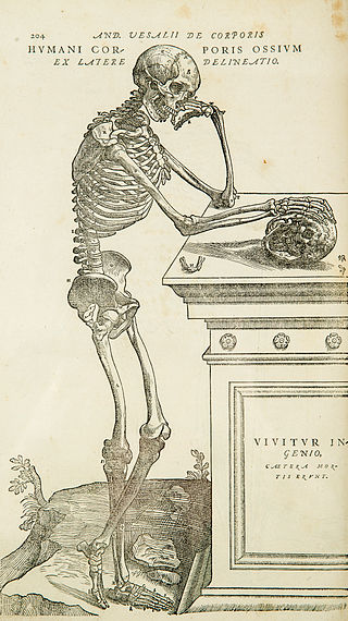
Anatomy is the branch of morphology concerned with the study of the internal structure of organisms and their parts. Anatomy is a branch of natural science that deals with the structural organization of living things. It is an old science, having its beginnings in prehistoric times. Anatomy is inherently tied to developmental biology, embryology, comparative anatomy, evolutionary biology, and phylogeny, as these are the processes by which anatomy is generated, both over immediate and long-term timescales. Anatomy and physiology, which study the structure and function of organisms and their parts respectively, make a natural pair of related disciplines, and are often studied together. Human anatomy is one of the essential basic sciences that are applied in medicine, and is often studied alongside physiology.
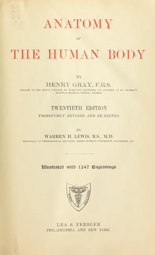
Gray's Anatomy is a reference book of human anatomy written by Henry Gray, illustrated by Henry Vandyke Carter and first published in London in 1858. It has had multiple revised editions and the current edition, the 42nd, remains a standard reference, often considered "the doctors' bible".

A joint or articulation is the connection made between bones, ossicles, or other hard structures in the body which link an animal's skeletal system into a functional whole. They are constructed to allow for different degrees and types of movement. Some joints, such as the knee, elbow, and shoulder, are self-lubricating, almost frictionless, and are able to withstand compression and maintain heavy loads while still executing smooth and precise movements. Other joints such as sutures between the bones of the skull permit very little movement in order to protect the brain and the sense organs. The connection between a tooth and the jawbone is also called a joint, and is described as a fibrous joint known as a gomphosis. Joints are classified both structurally and functionally.

Henry Gray was a British anatomist and surgeon most notable for publishing the book Gray's Anatomy. He was elected a Fellow of the Royal Society (FRS) at the age of 25.

In the human body, the lateral thoracic artery is a blood vessel that supplies oxygenated blood to approximately one-third of the lateral structures of the thorax and breast.
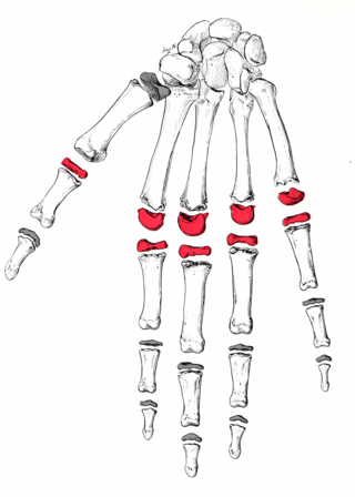
The metacarpophalangeal joints (MCP) are situated between the metacarpal bones and the proximal phalanges of the fingers. These joints are of the condyloid kind, formed by the reception of the rounded heads of the metacarpal bones into shallow cavities on the proximal ends of the proximal phalanges. Being condyloid, they allow the movements of flexion, extension, abduction, adduction and circumduction at the joint.

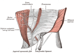
In human anatomy, the inguinal triangle is a region of the abdominal wall. It is also known by the eponym Hesselbach's triangle, after Franz Kaspar Hesselbach.

In human anatomy, the inferior epigastric vessels refers to the inferior epigastric artery and the inferior epigastric vein.

In human anatomy, the inferior epigastric artery is an artery that arises from the external iliac artery. It is accompanied by the inferior epigastric vein; inferiorly, these two inferior epigastric vessels together travel within the lateral umbilical fold The inferior epigastric artery then traverses the arcuate line of rectus sheath to enter the rectus sheath, then anastomoses with the superior epigastric artery within the rectus sheath.
In human anatomy, inferior epigastric vein are 1-2 veins accompanying the inferior epigastric artery. They drain into the external iliac vein just proximal to the inguinal ligament.
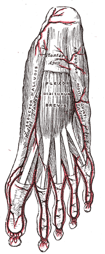
The abductor digiti minimi is a muscle which lies along the lateral (outer) border of the foot, and is in relation by its medial margin with the lateral plantar artery, vein and nerves.

The conjoint tendon is a sheath of connective tissue formed from the lower part of the common aponeurosis of the abdominal internal oblique muscle and the transversus abdominis muscle, joining the muscle to the pelvis. It forms the medial part of the posterior wall of the inguinal canal.
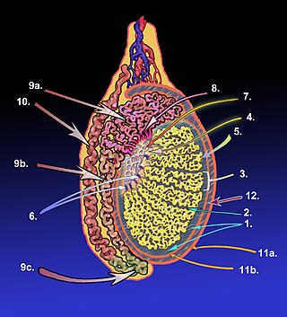
The tunica albuginea is a dense, blue-white layer of fibrous tissue surrounding the testis. It is the middle of three envelopes forming the capsule of the testis; it is deep to the visceral layer of tunica vaginalis, and superficial to the tunica vasculosa testis.

The retromandibular vein is a major vein of the face. It is formed within the parotid gland by the confluence of the maxillary vein, and superficial temporal vein. It descends in the gland and splits into two branches upon emerging from the gland. Its anterior branch then joins the (anterior) facial vein forming the common facial vein, while its posterior branch joins the posterior auricular vein forming the external jugular vein.

General visceral efferent fibers (GVE), visceral efferents or autonomic efferents are the efferent nerve fibers of the autonomic nervous system that provide motor innervation to smooth muscle, cardiac muscle, and glands through postganglionic varicosities.

The rectal venous plexus is the venous plexus surrounding the rectum. It consists of an internal and an external rectal plexus. It is drained by the superior, middle, and inferior rectal veins. It forms a portosystemic (portocaval) anastomosis. This allows rectally administered medications to bypass first pass metabolism.
The subcostal arteries, so named because they lie below the last ribs, constitute the lowest pair of branches derived from the thoracic aorta, and are in series with the intercostal arteries.

The femoral sheath is a funnel-shaped downward extension of abdominal fascia within which the femoral artery and femoral vein pass between the abdomen and the thigh. The femoral sheath is subdivided by two vertical partitions to form three compartments ; the medial compartment is known as the femoral canal and contains lymphatic vessels and a lymph node, whereas the intermediate canal and the lateral canal accommodate the femoral vein and the femoral artery (respectively). Some neurovascular structures perforate the femoral sheath. Topographically, the femoral sheath is contained within the femoral triangle.

The cribriform fascia, fascia cribrosa, also Hesselbach's fascia, is the portion of fascia covering the saphenous opening in the thigh. It is perforated by the great saphenous vein and by numerous blood and lymphatic vessels..

Anal columns are a number of vertical folds, produced by an infolding of the mucous membrane and some of the muscular tissue in the upper half of the lumen of the anal canal. They are named after Giovanni Battista Morgagni, who has several other eponyms named after him.