Deep perineal pouch

Last updated
Deep perineal pouch
Gray407.png
Coronal section of anterior part of pelvis, through the pubic arch. Seen from in front. (Deep perineal pouch not labeled, but is between the "superior layer" and "inferior layer" labeled at bottom left.)
Gray1156.png
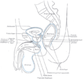
Vertical section of bladder, penis, and urethra. (Cowper's gland and membranous portion of urethra visible at center bottom.)
Details
Artery branches of internal pudendal artery
Vein branches of internal pudendal veins
Nerve branches of perineal nerve
Identifiers
Latin saccus profundus perinei or spatium perinei profundum
TA98 A09.5.03.001
TA2 2419
FMA 22061
Anatomical terminology

The deep perineal pouch (also deep perineal space) is the anatomic space enclosed in part by the perineum and located superior to the perineal membrane.

Contents

Structure

The deep perineal pouch is bordered inferiorly by the perineal membrane, also known as the inferior fascia of the urogenital diaphragm. It is bordered superiorly by the superior fascia of the urogenital diaphragm. [1] The deep pouch is now described as the region between the perineal membrane and the pelvic diaphragm. [2]

Contents

The deep perineal pouch contains:

Urogenital diaphragm

Older texts have asserted the existence of a "urogenital diaphragm", which was described as a layer of the pelvis that separates the deep perineal sac from the upper pelvis. The deep perineal pouch lies between the inferior fascia of the urogenital diaphragm and superior fascia of the urogenital diaphragm.

Additional images

See also

Related Research Articles

<span class="mw-page-title-main">Perineum</span> Region of the body including the perineal body and surrounding structures

The perineum in humans is the space between the anus and scrotum in the male or between the anus and vulva in the female. The perineum is the region of the body between the pubic symphysis and the coccyx, including the perineal body and surrounding structures. The perineal raphe is visible and pronounced to varying degrees. The perineum is an erogenous zone. This area is also known as the taint or gooch in American slang.

<span class="mw-page-title-main">Levator ani</span> Broad, thin muscle group, situated on either side of the pelvis

The levator ani is a broad, thin muscle group, situated on either side of the pelvis. It is formed from three muscle components: the pubococcygeus, the iliococcygeus, and the puborectalis.

<span class="mw-page-title-main">Bulbospongiosus muscle</span> Superficial muscle of the perineum

The bulbospongiosus muscle is one of the superficial muscles of the perineum. It has a slightly different origin, insertion and function in males and females. In males, it covers the bulb of the penis. In females, it covers the vestibular bulb.

<span class="mw-page-title-main">Pelvic floor</span> Anatomical structure

The pelvic floor or pelvic diaphragm is an anatomical location in the human body, which has an important role in urinary and anal continence, sexual function and support of the pelvic organs. The pelvic floor includes muscles, both skeletal and smooth, ligaments and fascia. and separates between the pelvic cavity from above, and the perineum from below. It is formed by the levator ani muscle and coccygeus muscle, and associated connective tissue.

<span class="mw-page-title-main">Urogenital diaphragm</span> Layer of the pelvis

Older texts have asserted the existence of a urogenital diaphragm, also called the triangular ligament, which was described as a layer of the pelvis that separates the deep perineal sac from the upper pelvis, lying between the inferior fascia of the urogenital diaphragm and superior fascia of the urogenital diaphragm.

<span class="mw-page-title-main">Anal canal</span> Functional segment of the large intestine

The anal canal is the part that connects the rectum to the anus, located below the level of the pelvic diaphragm. It is located within the anal triangle of the perineum, between the right and left ischioanal fossa. As the final functional segment of the bowel, it functions to regulate release of excrement by two muscular sphincter complexes. The anus is the aperture at the terminal portion of the anal canal.

<span class="mw-page-title-main">External anal sphincter</span> Flat plane of skeletal muscle fibers

The external anal sphincter is an oval tube of skeletal muscle fibers. Distally, it is adherent to the skin surrounding the margin of the anus. It exhibits a resting state of tonical contraction.

<span class="mw-page-title-main">External sphincter muscle of male urethra</span>

The external sphincter muscle of male urethra, also sphincter urethrae membranaceae, sphincter urethrae externus, surrounds the whole length of the membranous urethra, and is enclosed in the fascia of the urogenital diaphragm.

<span class="mw-page-title-main">Fascia of Colles</span>

The membranous layer of the superficial fascia of the perineum is the deeper layer of the superficial perineal fascia. It is thin, aponeurotic in structure, and of considerable strength, serving to bind down the muscles of the root of the penis. Colles' fascia emerges from the perineal membrane, which divides the base of the penis from the prostate. Colles' fascia emerges from the inferior side of the perineal membrane and continues along the ventral (inferior) penis without covering the scrotum. It separates the skin and subcutaneous fat from the superficial perineal pouch.

<span class="mw-page-title-main">Inferior anal nerves</span>

The inferior rectal nerves usually branch from the pudendal nerve but occasionally arises directly from the sacral plexus; they cross the ischiorectal fossa along with the inferior rectal artery and veins, toward the anal canal and the lower end of the rectum, and is distributed to the sphincter ani externus and to the integument (skin) around the anus.

<span class="mw-page-title-main">Transverse perineal muscles</span>

The transverse perineal muscles are the superficial and the deep transverse perineal muscles.

<span class="mw-page-title-main">Perineal membrane</span> Anatomical term

The perineal membrane is an anatomical term for a fibrous membrane in the perineum. The term "inferior fascia of urogenital diaphragm", used in older texts, is considered equivalent to the perineal membrane.

<span class="mw-page-title-main">Superficial perineal pouch</span>

The superficial perineal pouch is a compartment of the perineum.

<span class="mw-page-title-main">Urogenital triangle</span> Anterior part of the perineum

The urogenital triangle is the anterior part of the perineum. In female mammals, it contains the vagina and associated parts of the internal genitalia.

<span class="mw-page-title-main">Anal triangle</span> Posterior part of the perineum

The anal triangle is the posterior part of the perineum. It contains the anal canal.

<span class="mw-page-title-main">Membranous urethra</span> Narrowest part of the urethra

The membranous urethra or intermediate part of male urethra is the shortest, least dilatable, and, with the exception of the urinary meatus, the narrowest part of the urethra.

<span class="mw-page-title-main">Superior fascia of the urogenital diaphragm</span>

The superior fascia of the urogenital diaphragm is continuous with the obturator fascia and stretches across the pubic arch.

<span class="mw-page-title-main">Urethral sphincters</span> Muscles keeping urine in the bladder

The urethral sphincters are two muscles used to control the exit of urine in the urinary bladder through the urethra. The two muscles are either the male or female external urethral sphincter and the internal urethral sphincter. When either of these muscles contracts, the urethra is sealed shut.

<span class="mw-page-title-main">Root of penis</span> Internal portion of the human penis

In human male anatomy, the root of the penis is the internal and most proximal portion of the human penis that lies in the perineum. Unlike the pendulous body of the penis, which is suspended from the pubic symphysis, the root is attached to the pubic arch of the pelvis and is not visible externally. It is triradiate in form, consisting of three masses of erectile tissue; the two diverging crura, one on either side, and the median bulb of the penis or urethral bulb. Approximately one third of the penis is embedded in the pelvis and can be felt through the scrotum and in the perineum.

<span class="mw-page-title-main">Vaginal support structures</span> Structures that maintain the position of the vagina within the pelvic cavity

The vaginal support structures are those muscles, bones, ligaments, tendons, membranes and fascia, of the pelvic floor that maintain the position of the vagina within the pelvic cavity and allow the normal functioning of the vagina and other reproductive structures in the female. Defects or injuries to these support structures in the pelvic floor leads to pelvic organ prolapse. Anatomical and congenital variations of vaginal support structures can predispose a woman to further dysfunction and prolapse later in life. The urethra is part of the anterior wall of the vagina and damage to the support structures there can lead to incontinence and urinary retention.

References

  1. Netter, F., Atlas of Human Anatomy.4th Ed. Saunders, Philadelphia, 2006.
  2. Netter, F., Atlas of Human Anatomy.5th Ed. Saunders, Philadelphia, 2010.
  3. 1 2 Essential Clinical Anatomy – 4th Edition. p.255