
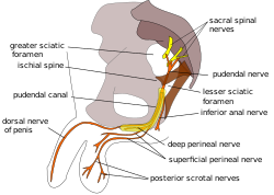
The pudendal nerve is the main nerve of the perineum. It is a mixed nerve and also conveys sympathetic autonomic fibers. It carries sensation from the external genitalia of both sexes and the skin around the anus and perineum, as well as the motor supply to various pelvic muscles, including the male or female external urethral sphincter and the external anal sphincter.

The levator ani is a broad, thin muscle group, situated on either side of the pelvis. It is formed from three muscle components: the pubococcygeus, the iliococcygeus, and the puborectalis.

The internal pudendal artery is one of the three pudendal arteries. It branches off the internal iliac artery, and provides blood to the external genitalia.

The pectineus muscle is a flat, quadrangular muscle, situated at the anterior (front) part of the upper and medial (inner) aspect of the thigh. The pectineus muscle is the most anterior adductor of the hip. The muscle's primary action is hip flexion; it also produces adduction and internal rotation of the hip.

The internal obturator muscle or obturator internus muscle originates on the medial surface of the obturator membrane, the ischium near the membrane, and the rim of the pubis.

In human anatomy, the inferior epigastric artery is an artery that arises from the external iliac artery. It is accompanied by the inferior epigastric vein; inferiorly, these two inferior epigastric vessels together travel within the lateral umbilical fold The inferior epigastric artery then traverses the arcuate line of rectus sheath to enter the rectus sheath, then anastomoses with the superior epigastric artery within the rectus sheath.

The obturator nerve in human anatomy arises from the ventral divisions of the second, third, and fourth lumbar nerves in the lumbar plexus; the branch from the third is the largest, while that from the second is often very small.

The sacrotuberous ligament is situated at the lower and back part of the pelvis. It is flat, and triangular in form; narrower in the middle than at the ends.

The superior gluteal artery is the terminal branch of the posterior division of the internal iliac artery. It exits the pelvis through the greater sciatic foramen before splitting into a superficial branch and a deep branch.
The inferior gluteal artery is a terminal branch of the anterior trunk of the internal iliac artery. It exits the pelvis through the greater sciatic foramen. It is distributed chiefly to the buttock and the back of the thigh.
The obturator artery is a branch of the internal iliac artery that passes antero-inferiorly on the lateral wall of the pelvis, to the upper part of the obturator foramen, and, escaping from the pelvic cavity through the obturator canal, it divides into an anterior branch and a posterior branch.

The inferior rectal artery is an artery that supplies blood to the lower third of the anal canal below the pectinate line.
The perineal nerve is a nerve of the pelvis. It arises from the pudendal nerve in the pudendal canal. It gives superficial branches to the skin, and a deep branch to muscles. It supplies the skin and muscles of the perineum. Its latency is tested with electrodes.

The ischioanal fossa is the fat-filled wedge-shaped space located lateral to the anal canal and inferior to the pelvic diaphragm. It is somewhat prismatic in shape, with its base directed to the surface of the perineum and its apex at the line of meeting of the obturator and anal fasciae.

The pelvic cavity is a body cavity that is bounded by the bones of the pelvis. Its oblique roof is the pelvic inlet. Its lower boundary is the pelvic floor.

The obturator fascia, or fascia of the internal obturator muscle, covers the pelvic surface of that muscle and is attached around the margin of its origin.
The inferior rectal nerves usually branch from the pudendal nerve but occasionally arises directly from the sacral plexus; they cross the ischiorectal fossa along with the inferior rectal artery and veins, toward the anal canal and the lower end of the rectum, and is distributed to the sphincter ani externus and to the integument (skin) around the anus.

The nerve to obturator internus is a mixed nerve providing motor innervation to the obturator internus muscle and gemellus superior muscle, and sensory innervation to the hip joint. It is a branch of the sacral plexus. It is one of the group of deep gluteal nerves.

The deep perineal pouch is the anatomic space enclosed in part by the perineum and located superior to the perineal membrane.

The anal triangle is the posterior part of the perineum. It contains the anus in mammals.
