Perineal nerve

Last updated
Perineal nerve
Pudendal nerve.svg
Pudendal nerve, its course through the lesser sciatic foramen, and branches, including inferior anal at bottom right.
Gray542.png
The superficial branches of the internal pudendal artery. (Perineal nerve visible but not labeled.)
Details
From pudendal nerve
Innervates perineum, scrotum or labia majora, superficial transverse perineal muscle, bulbospongiosus muscle, ischiocavernosus muscle, bulb of penis, levator ani, external anal sphincter
Identifiers
Latin nervi perineales
TA98 A14.2.07.039
TA2 6556
FMA 21866
Anatomical terms of neuroanatomy

The perineal nerve is a nerve of the pelvis. It arises from the pudendal nerve in the pudendal canal. It gives superficial branches to the skin, and a deep branch to muscles. It supplies the skin and muscles of the perineum. Its latency is tested with electrodes.

Contents

Structure

The perineal nerve is a branch of the pudendal nerve. [1] [2] It lies below the internal pudendal artery. It accompanies the perineal artery. It passes through the pudendal canal for around 2 or 3 cm. [1] [2] Whilst still in the canal, it divides into superficial branches and a deep branch. [1] The superficial branches of the perineal nerve become the posterior scrotal nerves in men, [3] and the posterior labial nerves in women. [1] The deep branch of the perineal nerve (also known as the "muscular" branch) travels to the muscles of the perineum. [1] Both of these are superficial to the dorsal nerve of the penis or the dorsal nerve of the clitoris. [4]

Function

The perineal nerve supplies the skin and muscles of the perineum. [1] The superficial branches supply sensation to the perineum, and the scrotum in men or the labia majora in women. [1] The deep branch supplies superficial transverse perineal muscle, the bulbospongiosus muscle, the ischiocavernosus muscle, the bulb of penis, levator ani, and the external anal sphincter. [1]

Clinical significance

The latency of the perineal nerve can be measured with electrodes. [5] It is used to test nerve function. [5]

Additional images

Related Research Articles

<span class="mw-page-title-main">Perineum</span> Region of the body between the genitals and anus

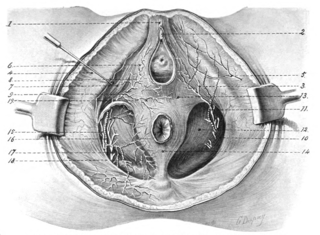
The perineum in mammals is the space between the anus and the genitals. The human perineum is between the anus and scrotum in the male or between the anus and vulva in the female. The perineum is the region of the body between the pubic symphysis and the coccyx, including the perineal body and surrounding structures. The perineal raphe is visible and pronounced to varying degrees. The perineum is an erogenous zone. This area is also known as the taint or gooch in American slang.

<span class="mw-page-title-main">Pudendal nerve</span> Main nerve of the perineum

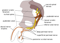
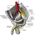
The pudendal nerve is the main nerve of the perineum. It is a mixed nerve and also conveys sympathetic autonomic fibers. It carries sensation from the external genitalia of both sexes and the skin around the anus and perineum, as well as the motor supply to various pelvic muscles, including the male or female external urethral sphincter and the external anal sphincter.

<span class="mw-page-title-main">Levator ani</span> Broad, thin muscle group, situated on either side of the pelvis

The levator ani is a broad, thin muscle group, situated on either side of the pelvis. It is formed from three muscle components: the pubococcygeus, the iliococcygeus, and the puborectalis.

<span class="mw-page-title-main">Bulbospongiosus muscle</span> Superficial muscle of the perineum

The bulbospongiosus muscles are one of the superficial muscles of the perineum. They have a slightly different origin, insertion and function in males and females. In males, these muscles cover the bulb of the penis, while in females, they cover the vestibular bulbs.

<span class="mw-page-title-main">Internal pudendal artery</span> Blood vessel supplying blood to the external genitalia

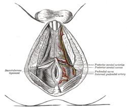
The internal pudendal artery is one of the three pudendal arteries. It branches off the internal iliac artery, and provides blood to the external genitalia.

<span class="mw-page-title-main">Obturator internus muscle</span> One of six small hip muscles in the lateral rotator group

The internal obturator muscle or obturator internus muscle originates on the medial surface of the obturator membrane, the ischium near the membrane, and the rim of the pubis.

<span class="mw-page-title-main">External anal sphincter</span> Flat plane of skeletal muscle fibers

The external anal sphincter is an oval tube of skeletal muscle fibers. Distally, it is adherent to the skin surrounding the margin of the anus. It exhibits a resting state of tonical contraction.

<span class="mw-page-title-main">Sacrotuberous ligament</span>

The sacrotuberous ligament is situated at the lower and back part of the pelvis. It is flat, and triangular in form; narrower in the middle than at the ends.

<span class="mw-page-title-main">Inferior rectal artery</span>

The inferior rectal artery is an artery that supplies blood to the lower third of the anal canal below the pectinate line.

<span class="mw-page-title-main">Internal pudendal veins</span> Set of veins in the pelvis enclosed by pudendal canal

The internal pudendal veins are a set of veins in the pelvis. They are the venae comitantes of the internal pudendal artery. Internal pudendal veins are enclosed by pudendal canal, with internal pudendal artery and pudendal nerve.

<span class="mw-page-title-main">Pudendal canal</span>

The pudendal canal is an anatomical structure formed by the obturator fascia lining the lateral wall of the ischioanal fossa. The internal pudendal artery and veins, and pudendal nerve pass through the pudendal canal, and the perineal nerve arises within it.

<span class="mw-page-title-main">Fascia of Colles</span>

The membranous layer of the superficial fascia of the perineum is the deeper layer of the superficial perineal fascia. It is thin, aponeurotic in structure, and of considerable strength, serving to bind down the muscles of the root of the penis. Colles' fascia emerges from the perineal membrane, which divides the base of the penis from the prostate. Colles' fascia emerges from the inferior side of the perineal membrane and continues along the ventral (inferior) penis without covering the scrotum. It separates the skin and subcutaneous fat from the superficial perineal pouch.

<span class="mw-page-title-main">Inferior anal nerves</span>

The inferior rectal nerves usually branch from the pudendal nerve but occasionally arises directly from the sacral plexus; they cross the ischiorectal fossa along with the inferior rectal artery and veins, toward the anal canal and the lower end of the rectum, and is distributed to the sphincter ani externus and to the integument (skin) around the anus.

<span class="mw-page-title-main">Dorsal artery of the penis</span> Artery of the penis

The dorsal artery of the penis is an artery on the top surface of the penis. It is a branch of the internal pudendal artery. It runs forward on the dorsum of the penis to the glans, where it divides into two branches to the glans penis and the foreskin (prepuce).

<span class="mw-page-title-main">Transverse perineal muscles</span>

The transverse perineal muscles are the superficial and the deep transverse perineal muscles.

<span class="mw-page-title-main">Deep perineal pouch</span> Anatomic space enclosed partly by the perineum

The deep perineal pouch is the anatomic space enclosed in part by the perineum and located superior to the perineal membrane.

<span class="mw-page-title-main">Anal triangle</span> Posterior part of the perineum

The anal triangle is the posterior part of the perineum. It contains the anal canal.

<span class="mw-page-title-main">Dorsal nerve of the clitoris</span>

The dorsal nerve of the clitoris is a nerve in females that branches off the pudendal nerve to innervate the clitoris. The nerve is important for female sexual pleasure, and it may play a role in clitoral erections.

The posterior labial nerves are branches of the pudendal nerve. They supply the female labia majora.

<span class="mw-page-title-main">Muscular branches of perineal nerve</span> Nerve of the perineum

The deep branch of the perineal nerve is a nerve of the perineum. It is a branch of the perineal nerve, from the pudendal nerve. It supplies the superficial transverse perineal muscle, bulbospongiosus muscle, ischiocavernosus muscle, the bulb of penis, levator ani, and the external anal sphincter.

References

PD-icon.svgThis article incorporates text in the public domain from page 968 of the 20th edition of Gray's Anatomy (1918)

  1. 1 2 3 4 5 6 7 8 Rea, Paul (2015). "3 - Lower Limb Nerve Supply". Essential Clinically Applied Anatomy of the Peripheral Nervous System in the Limbs. Academic Press. pp. 101–177. doi:10.1016/B978-0-12-803062-2.00003-6. ISBN   978-0-12-803062-2.
  2. 1 2 Vaida, George T.; Jain, Sudheer K. (2010). "6 - Anesthetic Complications in Urologic Surgery". Complications of Urologic Surgery - Prevention and Management. Saunders. pp. 57–82. doi:10.1016/B978-1-4160-4572-4.X0001-5. ISBN   978-1-4160-4572-4.
  3. Essential Clinical Anatomy. K.L. Moore & A.M. Agur. Lippincott, 2 ed. 2002. Page 263
  4. de Groat, William C.; Yoshimura, Naoki (2015). "5 - Anatomy and physiology of the lower urinary tract". Handbook of Clinical Neurology. Vol. 130. Elsevier. pp. 61–108. doi:10.1016/B978-0-444-63247-0.00005-5. ISBN   978-0-444-63247-0. ISSN   0072-9752. PMID   26003239.
  5. 1 2 Swash, Michael (2008). "33 - Sphincter Disorders and the Nervous System". Neurology and General Medicine (4th ed.). Churchill Livingstone. pp. 633–650. doi:10.1016/B978-044306707-5.50036-5. ISBN   978-0-443-06707-5.