Dorsal nerve of the penis

Last updated
Dorsal nerve of the penis
Pudendal nerve.svg
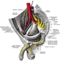
Pudendal nerve, its course through the lesser sciatic foramen, and branches, including dorsal nerve of the penis at bottom left.
Gray1155 a.png
Transverse section of the penis. (Dorsal nerve visible at top.)
Details
From Pudendal nerve
Innervates Penis
Identifiers
Latin nervus dorsalis penis
TA98 A14.2.07.042M
TA2 6563
FMA 21869
Anatomical terms of neuroanatomy

The dorsal nerve of the penis is the deepest of three divisions of the pudendal nerve; it accompanies the internal pudendal artery along the ramus of the ischium; it then runs forward along the margin of the inferior ramus of the pubis, between the superior and inferior layers of the fascia of the urogenital diaphragm.

Contents

Piercing the inferior layer it gives a branch to the corpus cavernosum penis, and passes forward, in company with the dorsal artery of the penis, between the layers of the suspensory ligament, on to the dorsum of the penis, and ends on the glans penis. [1]

It innervates the skin of the penis. In humans, it has 8290 ± 2553 axons, in 25–45 loosely packed nerve bundles, half of which are myelinated. The researches were conducted on volunteers that donated tissues after death, therefore some of the nerve endings could not have been counted due to the natural degeneration of tissues found in old people. [1]

See also

Related Research Articles

<span class="mw-page-title-main">Pudendal nerve</span> Main nerve of the perineum

The pudendal nerve is the main nerve of the perineum. It is a mixed nerve and also conveys sympathetic autonomic fibers. It carries sensation from the external genitalia of both sexes and the skin around the anus and perineum, as well as the motor supply to various pelvic muscles, including the male or female external urethral sphincter and the external anal sphincter.

<span class="mw-page-title-main">Bulbospongiosus muscle</span> Superficial muscle of the perineum

The bulbospongiosus muscles are a subgroup of the superficial muscles of the perineum. They have a slightly different origin, insertion and function in males and females. In males, these muscles cover the bulb of the penis, while in females, they cover the vestibular bulbs.

<span class="mw-page-title-main">Internal pudendal artery</span> Blood vessel supplying blood to the external genitalia

The internal pudendal artery is one of the three pudendal arteries. It branches off the internal iliac artery, and provides blood to the external genitalia.

<span class="mw-page-title-main">Inferior epigastric artery</span> Blood vessel

In human anatomy, the inferior epigastric artery is an artery that arises from the external iliac artery. It is accompanied by the inferior epigastric vein; inferiorly, these two inferior epigastric vessels together travel within the lateral umbilical fold The inferior epigastric artery then traverses the arcuate line of rectus sheath to enter the rectus sheath, then anastomoses with the superior epigastric artery within the rectus sheath.

<span class="mw-page-title-main">Sacrotuberous ligament</span>

The sacrotuberous ligament is situated at the lower and back part of the pelvis. It is flat, and triangular in form; narrower in the middle than at the ends.

<span class="mw-page-title-main">Inferior rectal artery</span>

The inferior rectal artery is an artery that supplies blood to the lower third of the anal canal below the pectinate line.

<span class="mw-page-title-main">Perineal nerve</span> Nerve of the perineum

The perineal nerve is a nerve of the pelvis. It arises from the pudendal nerve in the pudendal canal. It gives superficial branches to the skin, and a deep branch to muscles. It supplies the skin and muscles of the perineum. Its latency is tested with electrodes.

<span class="mw-page-title-main">Internal pudendal veins</span> Set of veins in the pelvis enclosed by pudendal canal

The internal pudendal veins are a set of veins in the pelvis. They are the venae comitantes of the internal pudendal artery. Internal pudendal veins are enclosed by pudendal canal, with internal pudendal artery and pudendal nerve.

<span class="mw-page-title-main">Celiac ganglia</span> Two large masses of nerve tissue in the upper abdomen

The celiac ganglia or coeliac ganglia are two large irregularly shaped masses of nerve tissue in the upper abdomen. Part of the sympathetic subdivision of the autonomic nervous system (ANS), the two celiac ganglia are the largest ganglia in the ANS, and they innervate most of the digestive tract.

<span class="mw-page-title-main">Thoracic splanchnic nerves</span>

Thoracic splanchnic nerves are splanchnic nerves that arise from the sympathetic trunk in the thorax and travel inferiorly to provide sympathetic supply to the abdomen. The nerves contain preganglionic sympathetic fibers and general visceral afferent fibers.

<span class="mw-page-title-main">Posterior scrotal nerves</span>

The posterior scrotal branches are two in number, medial and lateral. They are branches of the perineal nerve, which is itself a branch of the pudendal nerve. The pudendal nerve arises from spinal roots S2 through S4, travels through the pudendal canal on the fascia of the obturator internus muscle, and gives off the perineal nerve in the perineum. The major branch of the perineal nerve is the posterior scrotal/posterior labial.

<span class="mw-page-title-main">Inferior anal nerves</span>

The inferior rectal nerves usually branch from the pudendal nerve but occasionally arises directly from the sacral plexus; they cross the ischiorectal fossa along with the inferior rectal artery and veins, toward the anal canal and the lower end of the rectum, and is distributed to the sphincter ani externus and to the integument (skin) around the anus.

<span class="mw-page-title-main">Dorsal artery of the penis</span> Artery of the penis

The dorsal artery of the penis is an artery on the top surface of the penis. It is a branch of the internal pudendal artery. It runs forward on the dorsum of the penis to the glans, where it divides into two branches to the glans penis and the foreskin (prepuce).

<span class="mw-page-title-main">Medial umbilical ligament</span>

The medial umbilical ligament, cord of umbilical artery, or obliterated umbilical artery is a paired structure found in human anatomy. It is on the deep surface of the anterior abdominal wall, and is covered by the medial umbilical folds. It is different from the median umbilical ligament, a structure that represents the remnant of the embryonic urachus.

<span class="mw-page-title-main">Artery of bulb of penis</span>

The artery of bulb of penis is a short artery of large caliber which arises from the internal pudendal artery between the two layers of fascia of the urogenital diaphragm. It passes medialward, pierces the inferior fascia of the urogenital diaphragm and gives off branches which ramify in the bulb of the urethra and in the posterior part of the corpus spongiosum.

<span class="mw-page-title-main">Deep artery of the penis</span>

The deep artery of the penis is a branch of the internal pudendal artery that supplies the corpus spongiosum. The artery enters the crus of penis at its anterior extremity.

<span class="mw-page-title-main">Lumbosacral trunk</span> Nervous tissue that connects the lumbar plexus with the sacral plexus

The lumbosacral trunk is nervous tissue that connects the lumbar plexus with the sacral plexus. It is formed by the union of parts of the fourth and fifth lumbar nerves and descends to join the sacral plexus.

<span class="mw-page-title-main">Deep perineal pouch</span> Anatomic space enclosed partly by the perineum

The deep perineal pouch is the anatomic space enclosed in part by the perineum and located superior to the perineal membrane.

<span class="mw-page-title-main">Dorsal nerve of the clitoris</span>

The dorsal nerve of the clitoris is a nerve in females that branches off the pudendal nerve to innervate the clitoris. The nerve is important for female sexual pleasure, and it may play a role in clitoral erections.

<span class="mw-page-title-main">Muscular branches of perineal nerve</span> Nerve of the perineum

The deep branch of the perineal nerve is a nerve of the perineum. It is a branch of the perineal nerve, from the pudendal nerve. It supplies the superficial transverse perineal muscle, bulbospongiosus muscle, ischiocavernosus muscle, the bulb of penis, levator ani, and the external anal sphincter.

References

  1. 1 2 Tunçkol, Elçin; Purkart, Leopold; Eigen, Lennart; Vida, Imre; Brecht, Michael (2023). "Fiber counts and architecture of the human dorsal penile nerve". Scientific Reports. 13 (1). doi:10.1038/s41598-023-35030-w. ISSN   2045-2322. PMC   10232416 .

PD-icon.svgThis article incorporates text in the public domain from page 968 of the 20th edition of Gray's Anatomy (1918)