The perineum in mammals is the space between the anus and the genitals. The human perineum is between the anus and scrotum in the male or between the anus and vulva in the female. The perineum is the region of the body between the pubic symphysis and the coccyx, including the perineal body and surrounding structures. The perineal raphe is visible and pronounced to varying degrees. The perineum is an erogenous zone. This area is also known as the taint or gooch in American slang.

The bulbospongiosus muscles are a subgroup of the superficial muscles of the perineum. They have a slightly different origin, insertion and function in males and females. In males, these muscles cover the bulb of the penis, while in females, they cover the vestibular bulbs.

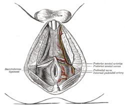
The internal pudendal artery is one of the three pudendal arteries. It branches off the internal iliac artery, and provides blood to the external genitalia.

The occipital artery is a branch of the external carotid artery that provides arterial supply to the back of the scalp, sternocleidomastoid muscles, and deep muscles of the back and neck.

The sacrotuberous ligament is situated at the lower and back part of the pelvis. It is flat, and triangular in form; narrower in the middle than at the ends.

The transverse cervical artery is an artery in the neck and a branch of the thyrocervical trunk, running at a higher level than the suprascapular artery.

The perineal nerve is a nerve of the pelvis. It arises from the pudendal nerve in the pudendal canal. It gives superficial branches to the skin, and a deep branch to muscles. It supplies the skin and muscles of the perineum. Its latency is tested with electrodes.

The membranous layer of the superficial fascia of the perineum is the deeper layer of the superficial perineal fascia. It is thin, aponeurotic in structure, and of considerable strength, serving to bind down the muscles of the root of the penis. Colles' fascia emerges from the perineal membrane, which divides the base of the penis from the prostate. Colles' fascia emerges from the inferior side of the perineal membrane and continues along the ventral (inferior) penis without covering the scrotum. It separates the skin and subcutaneous fat from the superficial perineal pouch.

The posterior scrotal branches are two in number, medial and lateral. They are branches of the perineal nerve, which is itself a branch of the pudendal nerve. The pudendal nerve arises from spinal roots S2 through S4, travels through the pudendal canal on the fascia of the obturator internus muscle, and gives off the perineal nerve in the perineum. The major branch of the perineal nerve is the posterior scrotal/posterior labial.

The transverse perineal muscles are the superficial and the deep transverse perineal muscles.

The deep external pudendal artery is one of the pudendal arteries that is more deeply seated than the superficial external pudendal artery, passes medially across the pectineus and the adductor longus muscles; it is covered by the fascia lata, which it pierces at the medial side of the thigh, and is distributed, in the male, to the integument of the scrotum and perineum, in the female to the labia majora; its branches anastomose with the scrotal or labial branches of the perineal artery.

The perineal membrane is an anatomical term for a fibrous membrane in the perineum. The term "inferior fascia of urogenital diaphragm", used in older texts, is considered equivalent to the perineal membrane.
The superficial perineal pouch is a compartment of the perineum.
The deep perineal pouch is the anatomic space enclosed in part by the perineum and located superior to the perineal membrane.

The urogenital triangle is the anterior part of the perineum. In female mammals, it contains the vagina and associated parts of the internal genitalia.

The posterior labial nerves are branches of the pudendal nerve. They supply the female labia majora.

The following outline is provided as an overview of and topical guide to human anatomy:
The deep branch of the perineal nerve is a nerve of the perineum. It is a branch of the perineal nerve, from the pudendal nerve. It supplies the superficial transverse perineal muscle, bulbospongiosus muscle, ischiocavernosus muscle, the bulb of penis, levator ani, and the external anal sphincter.

The pudendal arteries are a group of arteries which supply many of the muscles and organs of the pelvic cavity. The arteries include the internal pudendal artery, the superficial external pudendal artery, and the deep external pudendal artery.

The septum of scrotum or scrotal septum is an incomplete vertical wall (septum) that divides the scrotum into two compartments –each containing a single testis. It consists of flexible connective tissue and nonstriated muscle. The site of the median septum is apparent on the surface of the scrotum along a median longitudinal ridge called the scrotal raphe. The perineal raphe further extends forward to the undersurface of the penis and backward to the anal opening. The purpose of the median septum is to compartmentalize each testis in order to prevent friction or trauma.