Iliolumbar artery

Last updated
Iliolumbar artery
Gray585.png
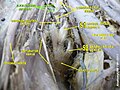
The veins of the right half of the male pelvis. (Iliolumbar artery not labeled, but Iliolumbar vein visible at center right.)
Details
Source Internal iliac artery
Branches Lumbar branches of iliolumbar artery
Vein Iliolumbar vein
Supplies Lumbar vertebrae, ilium
Identifiers
Latin arteria iliolumbalis
TA98 A12.2.15.002
TA2 4304
FMA 18845
Anatomical terminology

The iliolumbar artery is the first branch of the posterior trunk of the internal iliac artery.

Contents

Structure

The iliolumbar artery is the first branch of the posterior trunk of the internal iliac artery. [1] It turns upward behind the obturator nerve and the external iliac artery and vein, to the medial border of the psoas major muscle, behind which it divides into:

Anastomoses

Additional images

Related Research Articles

<span class="mw-page-title-main">Femoral triangle</span> Anatomical region of the thigh

The femoral triangle is an anatomical region of the upper third of the thigh. It is a subfascial space which appears as a triangular depression below the inguinal ligament when the thigh is flexed, abducted and laterally rotated.

<span class="mw-page-title-main">Deep femoral artery</span> Large branch of the femoral artery that supplies the thigh

The deep femoral artery also known as the deep artery of the thigh, or profunda femoris artery, is a large branch of the femoral artery. It travels more deeply ("profoundly") than the rest of the femoral artery. It gives rise to the lateral circumflex femoral artery and medial circumflex femoral artery, and the perforating arteries, terminating within the thigh.

<span class="mw-page-title-main">Pectineus muscle</span> Adductor of the thigh

The pectineus muscle is a flat, quadrangular muscle, situated at the anterior (front) part of the upper and medial (inner) aspect of the thigh. The pectineus muscle is the most anterior adductor of the hip. The muscle's primary action is hip flexion; it also produces adduction and internal rotation of the hip.

<span class="mw-page-title-main">External iliac artery</span> Arteries of the pelvis

The external iliac arteries are two major arteries which bifurcate off the common iliac arteries anterior to the sacroiliac joint of the pelvis.

<span class="mw-page-title-main">Internal iliac artery</span> Main artery of the pelvis

The internal iliac artery is the main artery of the pelvis.

<span class="mw-page-title-main">Inferior epigastric artery</span> Blood vessel

In human anatomy, the inferior epigastric artery is an artery that arises from the external iliac artery. It is accompanied by the inferior epigastric vein; inferiorly, these two inferior epigastric vessels together travel within the lateral umbilical fold The inferior epigastric artery then traverses the arcuate line of rectus sheath to enter the rectus sheath, then anastomoses with the superior epigastric artery within the rectus sheath.

<span class="mw-page-title-main">Femoral nerve</span> Long nerve down the thigh and inner leg

The femoral nerve is a nerve in the thigh that supplies skin on the upper thigh and inner leg, and the muscles that extend the knee. It is the largest branch of the lumbar plexus.

<span class="mw-page-title-main">Obturator nerve</span>

The obturator nerve in human anatomy arises from the ventral divisions of the second, third, and fourth lumbar nerves in the lumbar plexus; the branch from the third is the largest, while that from the second is often very small.

<span class="mw-page-title-main">Sacrotuberous ligament</span>

The sacrotuberous ligament is situated at the lower and back part of the pelvis. It is flat, and triangular in form; narrower in the middle than at the ends.

<span class="mw-page-title-main">Superior gluteal artery</span>

The superior gluteal artery is the terminal branch of the posterior division of the internal iliac artery. It exits the pelvis through the greater sciatic foramen before splitting into a superficial branch and a deep branch.

<span class="mw-page-title-main">Lateral sacral artery</span> Artery in the pelvis

The lateral sacral arteries is an artery in the pelvis that arises from the posterior division of the internal iliac artery. It later splits into two smaller branches, a superior and an inferior.

<span class="mw-page-title-main">Inferior gluteal artery</span>

The inferior gluteal artery is a terminal branch of the anterior trunk of the internal iliac artery. It exits the pelvis through the greater sciatic foramen. It is distributed chiefly to the buttock and the back of the thigh.

<span class="mw-page-title-main">Obturator artery</span>

The obturator artery is a branch of the internal iliac artery that passes antero-inferiorly on the lateral wall of the pelvis, to the upper part of the obturator foramen, and, escaping from the pelvic cavity through the obturator canal, it divides into an anterior branch and a posterior branch.

<span class="mw-page-title-main">Median sacral artery</span>

The median sacral artery is a small artery that arises posterior to the abdominal aorta and superior to its bifurcation.

<span class="mw-page-title-main">Lateral circumflex femoral artery</span>

The lateral circumflex femoral artery is an artery in the upper thigh. It is usually a branch of the profunda femoris artery, and produces three branches. It is mostly distributed to the muscles of the lateral thigh, supplying arterial blood to muscles of the knee extensor group.

<span class="mw-page-title-main">Lumbar arteries</span> Arteries within the lower back

The lumbar arteries are arteries located in the lower back or lumbar region. The lumbar arteries are in parallel with the intercostals.

The cruciate anastomosis is a circulatory anastomosis in the upper thigh formed by the inferior gluteal artery, the lateral and medial circumflex femoral arteries, the first perforating artery of the deep femoral artery, and the anastomotic branch of the posterior branch of the obturator artery.

In anatomy, arterial tree is used to refer to all arteries and/or the branching pattern of the arteries. This article regards the human arterial tree. Starting from the aorta:

<span class="mw-page-title-main">Deep circumflex iliac artery</span> Artery in the pelvis

The deep circumflex iliac artery is an artery in the pelvis that travels along the iliac crest of the pelvic bone.

<span class="mw-page-title-main">Outline of human anatomy</span> Overview of and topical guide to human anatomy

The following outline is provided as an overview of and topical guide to human anatomy:

References

PD-icon.svgThis article incorporates text in the public domain from page 621 of the 20th edition of Gray's Anatomy (1918)

  1. Paterson-Brown, Sara (2010-01-01), Bennett, Phillip; Williamson, Catherine (eds.), "Chapter Five - Applied anatomy", Basic Science in Obstetrics and Gynaecology (Fourth Edition), Churchill Livingstone, pp. 57–95, doi:10.1016/b978-0-443-10281-3.00009-9, ISBN   978-0-443-10281-3 , retrieved 2021-02-07
  2. Chait, A; Moltz, A; Nelson, JH (February 1968). "The collateral arterial circulation in the pelvis. An angiographic study". American Journal of Roentgenology. 102 (2): 392–400. doi:10.2214/ajr.102.2.392. PMID   5635691.