Testicular artery

Last updated
Testicular artery
Gray531.png
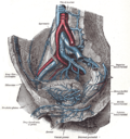
The abdominal aorta and its branches (internal spermatic vessels labeled at right)
Gray1149.png
Vertical section of the testis, to show the arrangement of the ducts (internal spermatic artery labeled vertically at center)
Details
Source Abdominal aorta
Vein Testicular vein
Identifiers
Latin arteria testicularis
TA98 A12.2.12.086M
TA2 4288
FMA 14758
Anatomical terminology

The testicular artery (the male gonadal artery, also called the internal spermatic arteries in older texts) is a branch of the abdominal aorta that supplies blood to the testicle. It is a paired artery, with one for each of the testicles.

Contents

It is the male equivalent of the ovarian artery. Because the testis is found in a different location than that of its female equivalent, it has a different course than the ovarian artery.

They are two slender vessels of considerable length, and arise from the front of the aorta a little below the renal arteries.

Each passes obliquely downward and lateralward behind the peritoneum, resting on the psoas major, the right lying in front of the inferior vena cava and behind the middle colic and ileocolic arteries and the terminal part of the ileum, the left behind the left colic and sigmoid arteries and the iliac colon.

Each crosses obliquely over the ureter and the lower part of the external iliac artery to reach the abdominal inguinal ring, through which it passes, and accompanies the other constituents of the spermatic cord along the inguinal canal to the scrotum, where it becomes tortuous, and divides into several branches.

Two or three of these accompany the ductus deferens, and supply the epididymis, anastomosing with the artery of the ductus deferens; others pierce the back part of the tunica albuginea, and supply the substance of the testicle.

The internal spermatic artery supplies one or two small branches to the ureter, and in the inguinal canal gives one or two twigs to the cremaster.

Additional images

See also

Related Research Articles

<span class="mw-page-title-main">Testicle</span> Internal organ in the male reproductive system

A testicle or testis is the male gonad in all bilaterians, including humans. It is homologous to the female ovary. The functions of the testicles are to produce both sperm and androgens, primarily testosterone. Testosterone release is controlled by the anterior pituitary luteinizing hormone, whereas sperm production is controlled both by the anterior pituitary follicle-stimulating hormone and gonadal testosterone.

<span class="mw-page-title-main">Vas deferens</span> Part of the male reproductive system of many vertebrates

The vas deferens, with the more modern name ductus deferens, is part of the male reproductive system of many vertebrates. The ducts transport sperm from the epididymis to the ejaculatory ducts in anticipation of ejaculation. The vas deferens is a partially coiled tube which exits the abdominal cavity through the inguinal canal.

<span class="mw-page-title-main">Spermatic cord</span> Structure in the human male reproductive system

The spermatic cord is the cord-like structure in males formed by the vas deferens and surrounding tissue that runs from the deep inguinal ring down to each testicle. Its serosal covering, the tunica vaginalis, is an extension of the peritoneum that passes through the transversalis fascia. Each testicle develops in the lower thoracic and upper lumbar region and migrates into the scrotum. During its descent it carries along with it the vas deferens, its vessels, nerves etc. There is one on each side.

<span class="mw-page-title-main">Cremaster muscle</span> Muscle covering the testicles and spermatic cord

The cremaster muscle is a paired structure made of thin layers of striated and smooth muscle that covers the testicles and the spermatic cord in human males. It consists of the lateral and medial parts. Cremaster is an involuntary muscle, responsible for the cremasteric reflex; a protective and physiologic superficial reflex of the testicles. The reflex raises and lowers the testicles in order to keep them protected. Along with the dartos muscle of the scrotum, it regulates testicular temperature, thus aiding the process of spermatogenesis.

<span class="mw-page-title-main">Inguinal canal</span> Human abdominal anatomy

The inguinal canal is a passage in the anterior abdominal wall on each side of the body, which in males, convey the spermatic cords and in females, the round ligament of the uterus. The inguinal canals are larger and more prominent in males.

<span class="mw-page-title-main">Abdominal aorta</span> Largest artery in the abdomen

In human anatomy, the abdominal aorta is the largest artery in the abdominal cavity. As part of the aorta, it is a direct continuation of the descending aorta.

<span class="mw-page-title-main">Male reproductive system</span> Reproductive system of the human male

The male reproductive system consists of a number of sex organs that play a role in the process of human reproduction. These organs are located on the outside of the body, and within the pelvis.

<span class="mw-page-title-main">External iliac vein</span> Blood vessels connecting the thigh veins to the pelvis

The external iliac veins are large veins that connect the femoral veins to the common iliac veins. Their origin is at the inferior margin of the inguinal ligaments and they terminate when they join the internal iliac veins.

<span class="mw-page-title-main">External iliac artery</span> Arteries of the pelvis

The external iliac arteries are two major arteries which bifurcate off the common iliac arteries anterior to the sacroiliac joint of the pelvis.

<span class="mw-page-title-main">Internal iliac artery</span> Main artery of the pelvis

The internal iliac artery is the main artery of the pelvis.

<span class="mw-page-title-main">Inferior epigastric artery</span> Blood vessel

In human anatomy, the inferior epigastric artery is an artery that arises from the external iliac artery. It is accompanied by the inferior epigastric vein; inferiorly, these two inferior epigastric vessels together travel within the lateral umbilical fold The inferior epigastric artery then traverses the arcuate line of rectus sheath to enter the rectus sheath, then anastomoses with the superior epigastric artery within the rectus sheath.

<span class="mw-page-title-main">Lumbar plexus</span> Web of nerves in the lower spine

The lumbar plexus is a web of nerves in the lumbar region of the body which forms part of the larger lumbosacral plexus. It is formed by the divisions of the first four lumbar nerves (L1-L4) and from contributions of the subcostal nerve (T12), which is the last thoracic nerve. Additionally, the ventral rami of the fourth lumbar nerve pass communicating branches, the lumbosacral trunk, to the sacral plexus. The nerves of the lumbar plexus pass in front of the hip joint and mainly support the anterior part of the thigh.

<span class="mw-page-title-main">Ovarian artery</span>

The ovarian artery is an artery that supplies oxygenated blood to the ovary in females. It arises from the abdominal aorta below the renal artery. It can be found within the suspensory ligament of the ovary, anterior to the ovarian vein and ureter.

<span class="mw-page-title-main">Testicular vein</span>

The testicular vein, the male gonadal vein, carries deoxygenated blood from its corresponding testis to the inferior vena cava or one of its tributaries. It is the male equivalent of the ovarian vein, and is the venous counterpart of the testicular artery.

<span class="mw-page-title-main">Cremasteric artery</span>

The cremasteric artery is a branch of the inferior epigastric artery which accompanies the spermatic cord to supply the cremaster muscle as well as other coverings of the spermatic cord in the male.

<span class="mw-page-title-main">Artery to the ductus deferens</span>

The artery to the ductus deferens is an artery in males that provides blood to the ductus deferens.

<span class="mw-page-title-main">Pampiniform plexus</span> Vein network in human males

The pampiniform plexus is a venous plexus – a network of many small veins found in the human male spermatic cord, and the suspensory ligament of the ovary. In the male, it is formed by the union of multiple testicular veins from the back of the testis and tributaries from the epididymis.

<span class="mw-page-title-main">Outline of human anatomy</span> Overview of and topical guide to human anatomy

The following outline is provided as an overview of and topical guide to human anatomy:

<span class="mw-page-title-main">Scrotum</span> Sac of skin that protects the testicles

In most terrestrial mammals, the scrotum or scrotal sac is a part of the external male genitalia located at the base of the penis that consists of a suspended dual-chambered sac of skin and smooth muscle. The scrotum contains the external spermatic fascia, testicles, epididymis, and ductus deferens. It is a distention of the perineum and carries some abdominal tissues into its cavity including the testicular artery, testicular vein, and pampiniform plexus. The perineal raphe is a small, vertical, slightly raised ridge of scrotal skin under which is found the scrotal septum. It appears as a thin longitudinal line that runs front to back over the entire scrotum. In humans, the scrotum becomes covered with pubic hair at puberty. The scrotum will usually tighten during penile erection and when exposed to cold temperatures. One testis is typically lower than the other to avoid compression in the event of an impact.

<span class="mw-page-title-main">Scrotal ultrasound</span> Medical ultrasound examination of the scrotum.

Scrotalultrasound is a medical ultrasound examination of the scrotum. It is used in the evaluation of testicular pain, and can help identify solid masses.

References

PD-icon.svgThis article incorporates text in the public domain from page 611 of the 20th edition of Gray's Anatomy (1918)