In many Animalia, including humans, an ileocolic structure or problem is something that concerns the region of the gastrointestinal tract from the ileum to the colon. In Animalia that have ceca, the ileocecal region is a subset of the ileocolic region, and the entire range can also be described as ileocecocolic, whereas in some Animalia, the ileocolic region contains no cecum, as the ileum joins the colon directly.
Things that are ileocolic, ileocecal, or both include the following:
This article includes a list of related items that share the same name (or similar names). If an internal link incorrectly led you here, you may wish to change the link to point directly to the intended article.
Related Research Articles
The cecum or caecum is a pouch within the peritoneum that is considered to be the beginning of the large intestine. It is typically located on the right side of the body. The word cecum stems from the Latin caecus meaning blind.
The ileum is the final section of the small intestine in most higher vertebrates, including mammals, reptiles, and birds. In fish, the divisions of the small intestine are not as clear and the terms posterior intestine or distal intestine may be used instead of ileum. Its main function is to absorb vitamin B12, bile salts, and whatever products of digestion that were not absorbed by the jejunum.
Enteric duplication cysts, sometimes simply called duplication cysts, are rare congenital malformations of the gastrointestinal tract. They most frequently occur in the small intestine, particularly the ileum, but can occur anywhere along the gastrointestinal tract. They may be cystic or tubular in conformation.
Intussusception is a medical condition in which a part of the intestine folds into the section immediately ahead of it. It typically involves the small bowel and less commonly the large bowel. Symptoms include abdominal pain which may come and go, vomiting, abdominal bloating, and bloody stool. It often results in a small bowel obstruction. Other complications may include peritonitis or bowel perforation.
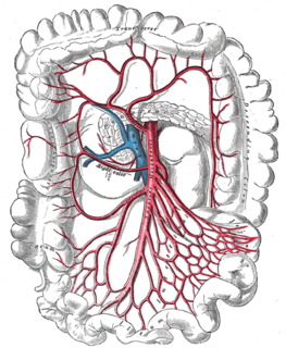
In human anatomy, the superior mesenteric artery (SMA) is an artery which arises from the anterior surface of the abdominal aorta, just inferior to the origin of the celiac trunk, and supplies blood to the intestine from the lower part of the duodenum through two-thirds of the transverse colon, as well as the pancreas.
Lower gastrointestinal bleeding, commonly abbreviated LGIB, is any form of gastrointestinal bleeding in the lower gastrointestinal tract. LGIB is a common reason for seeking medical attention at a hospital's emergency department. LGIB accounts for 30–40% of all gastrointestinal bleeding and is less common than upper gastrointestinal bleeding (UGIB). It is estimated that UGIB accounts for 100–200 per 100,000 cases versus 20–27 per 100,000 cases for LGIB. Approximately 85% of lower gastrointestinal bleeding involves the colon, 10% are from bleeds that are actually upper gastrointestinal bleeds, and 3–5% involve the small intestines.
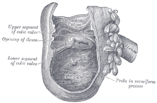
The ileocecal valve is a sphincter muscle valve that separates the small intestine and the large intestine. Its critical function is to limit the reflux of colonic contents into the ileum. Approximately two liters of fluid enters the colon daily through the ileocecal valve.
The right colic artery is an artery of the abdomen. It is a branch of the superior mesenteric artery. It supplies the ascending colon. It may be removed in a right hemicolectomy.
The ileocolic artery is the lowest branch arising from the concavity of the superior mesenteric artery.
The appendicular artery, also known as appendiceal artery, commonly arises from the terminal branch of the ileocolic artery, or less commonly from the posterior cecal artery or an ileal artery. It descends behind the termination of the ileum and enters the mesoappendix of the vermiform appendix. It runs near the free margin of the mesoappendix and ends in branches which supply the appendix.
The testicular artery is a branch of the abdominal aorta that supplies blood to the testis. It is a paired artery, with one for each of the testes.
The bow and arrow sign is an endoscopic sign for determining the location of the ileocecal valve during colonoscopy. Identifying the ileocecal valve in a colonoscopy is important, as it indicates that the entire colon has been visualized.
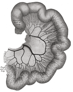
The arterial arcades are a series of anastomosing arterial arches between the arterial branches of the jejunum and ileum.
Colic artery may refer to the:
The ileocecal fold is an anatomical structure of the human abdomen formed by a layer of peritoneum between the ileum and cecum. The upper border of the ileocecal fold is fixed to the ileum opposite its mesenteric attachment, and the lower border passes over the ileocecal junction to join the mesentery of the appendix. Behind the ileocecal fold is the inferior ileocecal fossa.
The ileocolic vein is a vein which drains the ileum, colon, and cecum.
The superior mesenteric lymph nodes may be divided into three principal groups:
Cecal bascule is a cause of large bowel obstruction where there is folding of the cecum anteriorly over the ascending colon. It is one of two types of cecal volvulus, the other being axial ileocolic. It is caused by rotational torsion of the cecum or ascending colon along its own axis. In cecal bascule, the base of the cecum folds anteriorly over the ascending colon, creating a flap-valve, obstructing emptying of the cecum. The condition can be complicated by necrosis or organ perforation before the diagnosis is made, particularly if the ileocecal valve is competent, preventing retrograde decompression of the cecum into the ileum.
The gastroileal reflex is one of the three extrinsic reflexes of the gastrointestinal tract, the other two being the gastrocolic reflex and the enterogastric reflex. The gastroileal reflex is stimulated by the presence of food in the stomach and gastric peristalsis. Initiation of the reflex causes peristalsis in the ileum and the opening of the ileocecal valve. This in turn stimulates colonic peristalsis and an urge to defecate.
This page is based on this Wikipedia article Text is available under the CC BY-SA 4.0 license; additional terms may apply. Images, videos and audio are available under their respective licenses.