Femoral artery

Last updated

Femoral artery
Sobo 1909 573-574.png
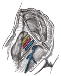
Thigh with and without the sartorius muscle, revealing the femoral artery and vein underneath
Details
Source External iliac artery
Branches Superficial epigastric artery, superficial iliac circumflex, superficial external pudendal, deep external pudendal, deep femoral artery,continues as popliteal artery
Vein Femoral vein
Supplies Anterior compartment of thigh
Identifiers
Latin arteria femoralis
MeSH D005263
TA98 A12.2.16.010
TA2 4674
FMA 70248
Anatomical terminology

The femoral artery is a large artery in the thigh and the main arterial supply to the thigh and leg. The femoral artery gives off the deep femoral artery and descends along the anteromedial part of the thigh in the femoral triangle. It enters and passes through the adductor canal, and becomes the popliteal artery as it passes through the adductor hiatus in the adductor magnus near the junction of the middle and distal thirds of the thigh. [1]

Contents

The femoral artery proximal to the origin of the deep femoral artery is referred to as the common femoral artery, whereas the femoral artery distal to this origin is referred to as the superficial femoral artery. [2]

Structure

Femoral artery showing common and superficial arteries, in common usage but not listed in TA Femoral-artery-grays-illustrations.jpg
Femoral artery showing common and superficial arteries, in common usage but not listed in TA

The femoral artery represents the continuation of the external iliac artery beyond the inguinal ligament underneath which the vessel passes [2] to enter the thigh. [3] The vessel passes under the inguinal ligament just medial of the midpoint of this ligament, [2] midway between the anterior superior iliac spine and the symphysis pubis (mid-inguinal point).[ citation needed ]

In common usage, in clinical practice including angiology and vascular surgery, the femoral artery includes the common femoral artery, and the superficial femoral artery however, the Terminologia Anatomica (TA) only lists the femoral artery. (The TA is the international standard for human anatomical terminology developed by the Federative International Programme on Anatomical Terminology). [4]

Relations

The relations of the femoral artery are as follows:

Branches

Schema of arteries of the thigh, including femoral artery and branches. Thigh arteries schema.svg
Schema of arteries of the thigh, including femoral artery and branches.

Common femoral artery

Superficial femoral artery

Clinical significance

Clinical examination

The site for optimally palpating the femoral pulse is in the inner thigh, at the mid-inguinal point, halfway between the pubic symphysis and anterior superior iliac spine. Presence of a femoral pulse indicates a systolic blood pressure of more than 50 mmHg. [15]

Vascular access

Femoral artery is the frequent site of access in angiography. As the pulsation of the common femoral artery can often be palpated through the skin; and the site of maximum pulsation is used as a point of puncture for catheter access. [5] From here, wires and catheters can be directed anywhere in the arterial system for intervention or diagnostics, including the heart, brain, kidneys, arms and legs. The direction of the needle in the femoral artery can be against blood flow (retro-grade), for intervention and diagnostic towards the heart and opposite leg, or with the flow (ante-grade or ipsi-lateral) for diagnostics and intervention on the same leg. Access in either the left or right femoral artery is possible and depends on the type of intervention or diagnostic.[ citation needed ]

To image the lower limb vascular anatomy, the common femoral artery (CFA) is chosen as the site of entry. However, CFA entry can only be assessed by retrograde puncture. Therefore, a catheter is advanced retrogradely through the contralateral common femoral artery into common iliac artery, crossing the midline into ipsilateral CFA. The SFA can then be assessed by antegrade puncture. [16]

The femoral artery can be used to draw arterial blood when the blood pressure is so low that the radial or brachial arteries cannot be located.

Peripheral arterial disease

The femoral artery is susceptible to peripheral arterial disease. [17] When it is blocked through atherosclerosis, percutaneous intervention with access from the opposite femoral may be needed. Endarterectomy, a surgical cut down and removal of the plaque of the femoral artery is also common. If the femoral artery has to be ligated surgically to treat a popliteal aneurysm, blood can still reach the popliteal artery distal to the ligation via the genicular anastomosis. However, if flow in the femoral artery of a normal leg is suddenly disrupted, blood flow distally is rarely sufficient. The reason for this is the fact that the genicular anastomosis is only present in a minority of individuals and is always undeveloped when disease in the femoral artery is absent. [18]

See also

Related Research Articles

<span class="mw-page-title-main">Human leg</span> Lower extremity or limb of the human body (foot, lower leg, thigh and hip)

The human leg is the entire lower limb of the human body, including the foot, thigh or sometimes even the hip or buttock region. The major bones of the leg are the femur, tibia, and adjacent fibula. The thigh is between the hip and knee, while the calf (rear) and shin (front) are between the knee and foot.

<span class="mw-page-title-main">Thigh</span> Area between the pelvis and the knee; upper leg

In human anatomy, the thigh is the area between the hip (pelvis) and the knee. Anatomically, it is part of the lower limb.

<span class="mw-page-title-main">Femoral triangle</span> Anatomical region of the thigh

The femoral triangle is an anatomical region of the upper third of the thigh. It is a subfascial space which appears as a triangular depression below the inguinal ligament when the thigh is flexed, abducted and laterally rotated.

<span class="mw-page-title-main">Great saphenous vein</span> Large, subcutaneous, superficial vein of the leg

The great saphenous vein(GSV), or long saphenous vein; ) is a large, subcutaneous, superficial vein of the leg. It is the longest vein in the body, running along the length of the lower limb, returning blood from the foot, leg and thigh to the deep femoral vein at the femoral triangle.

<span class="mw-page-title-main">Popliteal artery</span> Continuation of the femoral artery that supplies the lower leg

The popliteal artery is a deeply placed continuation of the femoral artery opening in the distal portion of the adductor magnus muscle. It courses through the popliteal fossa and ends at the lower border of the popliteus muscle, where it branches into the anterior and posterior tibial arteries.

<span class="mw-page-title-main">Inguinal lymph nodes</span> Lymph nodes in the human groin

Inguinal lymph nodes are lymph nodes in the groin. They are situated in the femoral triangle of the inguinal region. They are subdivided into two groups: the superficial inguinal lymph nodes and deep inguinal lymph nodes.

<span class="mw-page-title-main">Groin</span> The two creases at the junction of the torso with the thighs, on either side of the pubic area

In human anatomy, the groin also known as the inguinal region or iliac region, is the junctional area between the torso and the thigh. The groin is at the front of the body on either side of the pubic tubercle, where the lower part of the abdominal wall meets the thigh. A fold or crease is formed at this junction known as the inguinal groove, or crease. This is also the area of the medial compartment of the thigh that contains attachments of the adductor muscles of the hip or the groin muscles. The groin is the common site for a hernia.

<span class="mw-page-title-main">Femoral vein</span> Large blood vessel in the leg

In the human body, the femoral vein is the vein that accompanies the femoral artery in the femoral sheath. It is a deep vein that begins at the adductor hiatus as the continuation of the popliteal vein. The great saphenous vein, and the deep femoral vein drain into the femoral vein in the femoral triangle when it becomes known as the common femoral vein. It ends at the inferior margin of the inguinal ligament where it becomes the external iliac vein. Its major tributaries are the deep femoral vein, and the great saphenous vein. The femoral vein contains valves.

<span class="mw-page-title-main">External iliac artery</span> Arteries of the pelvis

The external iliac arteries are two major arteries which bifurcate off the common iliac arteries anterior to the sacroiliac joint of the pelvis.

<span class="mw-page-title-main">Adductor magnus muscle</span> Muscle in the thigh

The adductor magnus is a large triangular muscle, situated on the medial side of the thigh.

<span class="mw-page-title-main">Gracilis muscle</span> Most superficial muscle on the medial side of the thigh

The gracilis muscle is the most superficial muscle on the medial side of the thigh. It is thin and flattened, broad above, narrow and tapering below.

<span class="mw-page-title-main">Iliopsoas</span> Joined psoas and the iliacus muscles.

The iliopsoas muscle refers to the joined psoas major and the iliacus muscles. The two muscles are separate in the abdomen, but usually merge in the thigh. They are usually given the common name iliopsoas. The iliopsoas muscle joins to the femur at the lesser trochanter. It acts as the strongest flexor of the hip.

<span class="mw-page-title-main">Internal iliac artery</span> Main artery of the pelvis

The internal iliac artery is the main artery of the pelvis.

<span class="mw-page-title-main">Popliteal fossa</span> Depression at back of knee joint

The popliteal fossa is a shallow depression located at the back of the knee joint. The bones of the popliteal fossa are the femur and the tibia. Like other flexion surfaces of large joints, it is an area where blood vessels and nerves pass relatively superficially, and with an increased number of lymph nodes.

<span class="mw-page-title-main">Lateral cutaneous nerve of thigh</span> Nerve of the thigh

The lateral cutaneous nerve of the thigh is a cutaneous nerve of the thigh. It originates from the dorsal divisions of the second and third lumbar nerves from of lumbar plexus. It passes under the inguinal ligament to reach the thigh. It supplies sensation to the skin on the lateral part of the thigh by an anterior branch and a posterior branch.

In anatomy, arterial tree is used to refer to all arteries and/or the branching pattern of the arteries. This article regards the human arterial tree. Starting from the aorta:

<span class="mw-page-title-main">Superficial external pudendal artery</span>

The superficial external pudendal artery is one of the three pudendal arteries. It arises from the medial side of the femoral artery, close to the superficial epigastric artery and superficial iliac circumflex artery.

<span class="mw-page-title-main">Adductor hiatus</span> Gap between the adductor magnus muscle and the femur

In human anatomy, the adductor hiatus also known as hiatus magnus is a hiatus (gap) between the adductor magnus muscle and the femur that allows the passage of the femoral vessels from the anterior thigh to the posterior thigh and then the popliteal fossa. It is the termination of the adductor canal and lies about 8–13.5 cm. superior to the adductor tubercle.

<span class="mw-page-title-main">Patellar network</span> Network of blood vessels around the knee

The patellar network is an intricate network of blood vessels around and above the patella, and on the contiguous ends of the femur and tibia, forming a superficial and a deep plexus.

<span class="mw-page-title-main">Outline of human anatomy</span> Overview of and topical guide to human anatomy

The following outline is provided as an overview of and topical guide to human anatomy:

References

  1. Schulte, Erik; Schumacher, Udo (2006). "Arterial Supply to the Thigh". In Ross, Lawrence M.; Lamperti, Edward D. (eds.). Thieme Atlas of Anatomy: General Anatomy and Musculoskeletal System. Thieme. p. 490. ISBN   978-3-13-142081-7.
  2. 1 2 3 Swift, Hilary; Bordoni, Bruno (2022), "Anatomy, Bony Pelvis and Lower Limb, Femoral Artery", StatPearls, Treasure Island (FL): StatPearls Publishing, PMID   30855850 , retrieved January 11, 2023
  3. Jacob, S. (January 1, 2008), Jacob, S. (ed.), "Chapter 6 - Lower limb", Human Anatomy, Churchill Livingstone, pp. 135–179, doi:10.1016/b978-0-443-10373-5.50009-9, ISBN   978-0-443-10373-5 , retrieved January 18, 2021
  4. Kachlik D, Musil V, Blankova A, Marvanova Z, Miletin J, Trachtova D; et al. (2021). "A plea for extension of the anatomical nomenclature: Vessels". Bosn J Basic Med Sci. 21 (2): 208–220. doi:10.17305/bjbms.2020.5256. PMC   7982069 . PMID   33259774.{{cite journal}}: CS1 maint: multiple names: authors list (link)
  5. 1 2 van den Berg, Jos C (January 2013). "Optimal Technique for Common Femoral Artery Access". Endovascular Today. Archived from the original on August 6, 2021. Retrieved August 6, 2021.
  6. Sandgren T, Sonesson B, Ahlgren R, Länne T (1999). "The diameter of the common femoral artery in healthy human: influence of sex, age, and body size". J Vasc Surg. 29 (3): 503–10. doi: 10.1016/s0741-5214(99)70279-x . PMID   10069915.{{cite journal}}: CS1 maint: multiple names: authors list (link)
  7. 1 2 3 Trani C, Russo G, Aurigemma C, Burzotta F (2019). "The conundrum of endovascular common femoral artery treatment: a case report of lithoplasty as a viable solution". Eur Heart J Case Rep. 3 (3): ytz122. doi:10.1093/ehjcr/ytz122. PMC   6764558 . PMID   31660495.{{cite journal}}: CS1 maint: multiple names: authors list (link)
  8. Snell, Richard S. (2008). Clinical Anatomy By Regions (8 ed.). Baltimore: Lippincott Williams & Wilkins. pp. 581–582. ISBN   978-0-7817-6404-9.
  9. Bundens, WP; Bergan, JJ; Halasz, NA; Murray, J; Drehobl, M (1995). "The superficial femoral vein. A potentially lethal misnomer". JAMA. 274 (16): 1296–8. doi:10.1001/jama.1995.03530160048032. PMID   7563535.
  10. Hammond, I (2003). "The superficial femoral vein". Radiology. 229 (2): 604, discussion 604-6. doi:10.1148/radiol.2292030418. PMID   14595157.
  11. Kitchens CS (2011). "How I treat superficial venous thrombosis". Blood. 117 (1): 39–44. doi: 10.1182/blood-2010-05-286690 . PMID   20980677.
  12. Thiagarajah R, Venkatanarasimha N, Freeman S (2011). "Use of the term "superficial femoral vein" in ultrasound". J Clin Ultrasound. 39 (1): 32–34. doi:10.1002/jcu.20747. PMID   20957733. S2CID   23215861.
  13. Amarnath C and Hemant Patel (2023). Comprehensive Textbook of Clinical Radiology - Volume III: Chest and Cardiovascular system. Elsevier Health Sciences. ISBN   9788131263617. Page 1072
  14. 1 2 3 4 5 Ryan, Stephanie (2011). "Chapter 8". Anatomy for diagnostic imaging (Third ed.). Elsevier Ltd. p. 306. ISBN   9780702029714.
  15. Deakin, Charles D.; Low, J. Lorraine (September 2000). "Accuracy of the advanced trauma life support guidelines for predicting systolic blood pressure using carotid, femoral, and radial pulses: observational study". BMJ . 321 (7262): 673–4. doi:10.1136/bmj.321.7262.673. PMC   27481 . PMID   10987771.
  16. Berman, Hl; Katz, Sg; Tihansky, Dp (September 1986). "Guided direct antegrade puncture of the superficial femoral artery". American Journal of Roentgenology. 147 (3): 632–634. doi:10.2214/ajr.147.3.632. ISSN   0361-803X. PMID   2943146.
  17. MacPherson, D. S.; Evans, D. H.; Bell, P. R. F. (January 1984). "Common femoral artery Doppler wave-forms: a comparison of three methods of objective analysis with direct pressure measurements". British Journal of Surgery . 71 (1): 46–9. doi:10.1002/bjs.1800710114. PMID   6689970. S2CID   30352039.
  18. Sabalbal, M.; Johnson, M.; McAlister, V. (September 2013). "Absence of the genicular arterial anastomosis as generally depicted in textbooks". Annals of the Royal College of Surgeons of England . 95 (6): 405–9. doi:10.1308/003588413X13629960046831. PMC   4188287 . PMID   24025288.

Additional images