
The gluteus minimus, or glutæus minimus, the smallest of the three gluteal muscles, is situated immediately beneath the gluteus medius.

In human anatomy, the sacral plexus is a nerve plexus which provides motor and sensory nerves for the posterior thigh, most of the lower leg and foot, and part of the pelvis. It is part of the lumbosacral plexus and emerges from the lumbar vertebrae and sacral vertebrae (L4-S4). A sacral plexopathy is a disorder affecting the nerves of the sacral plexus, usually caused by trauma, nerve compression, vascular disease, or infection. Symptoms may include pain, loss of motor control, and sensory deficits.

The internal iliac artery is the main artery of the pelvis.
The inferior gluteal nerve is the main motor neuron that innervates the gluteus maximus muscle. It is responsible for the movement of the gluteus maximus in activities requiring the hip to extend the thigh, such as climbing stairs. Injury to this nerve is rare but often occurs as a complication of posterior approach to the hip during hip replacement. When damaged, one would develop gluteus maximus lurch, which is a gait abnormality which causes the individual to 'lurch' backwards to compensate lack in hip extension.
The superior gluteal nerve is a mixed nerve of the sacral plexus that originates in the pelvis. It provides motor innervation to the gluteus medius, gluteus minimus, tensor fasciae latae, and piriformis muscles; it also has a cutaneous branch.

The sacrotuberous ligament is situated at the lower and back part of the pelvis. It is flat, and triangular in form; narrower in the middle than at the ends.
The sacrospinous ligament is a thin, triangular ligament in the human pelvis. The base of the ligament is attached to the outer edge of the sacrum and coccyx, and the tip of the ligament attaches to the spine of the ischium, a bony protuberance on the human pelvis. Its fibres are intermingled with the sacrotuberous ligament.

The uterine artery is an artery that supplies blood to the uterus in females.

The iliolumbar artery is the first branch of the posterior trunk of the internal iliac artery.

The superior gluteal artery is the terminal branch of the posterior division of the internal iliac artery. It exits the pelvis through the greater sciatic foramen before splitting into a superficial branch and a deep branch.

The lateral sacral arteries is an artery in the pelvis that arises from the posterior division of the internal iliac artery. It later splits into two smaller branches, a superior and an inferior.
The inferior gluteal artery is a terminal branch of the anterior trunk of the internal iliac artery. It exits the pelvis through the greater sciatic foramen. It is distributed chiefly to the buttock and the back of the thigh.
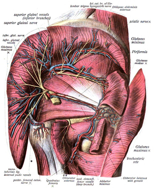
The inferior gluteal veins are venae comitantes of the inferior gluteal artery. They commence in the superior/proximal posterior thigh. They enter the pelvis through the lower part of the greater sciatic foramen. They converge to form a single vessel before emptying into the distal portion of the internal iliac vein.
The superior gluteal veins are venæ comitantes of the superior gluteal artery. They receive tributaries from the buttock corresponding with the branches of the artery. They enter the pelvis through the greater sciatic foramen, superior to the piriformis. They drain into internal iliac vein.
The cruciate anastomosis is a circulatory anastomosis in the upper thigh formed by the inferior gluteal artery, the lateral and medial circumflex femoral arteries, the first perforating artery of the deep femoral artery, and the anastomotic branch of the posterior branch of the obturator artery.

The greater sciatic notch is a notch in the ilium, one of the bones that make up the human pelvis. It lies between the posterior inferior iliac spine (above), and the ischial spine (below). The sacrospinous ligament changes this notch into an opening, the greater sciatic foramen.

The deep circumflex iliac artery is an artery in the pelvis that travels along the iliac crest of the pelvic bone.

The accompanying artery of ischiadic nerve is a long, slender artery in the thigh. It branches of the inferior gluteal artery. It accompanies the sciatic nerve for a short distance. It then penetrates it, and runs in its substance to the lower part of the thigh.

The superficial branch of medial circumflex femoral artery appears between the quadratus femoris and upper border of the adductor magnus, and anastomoses with the inferior gluteal artery, lateral femoral circumflex artery, and first of the perforating arteries of the deep femoral artery.

The gemelli muscles are the inferior gemellus muscle and the superior gemellus muscle, two small accessory fasciculi to the tendon of the internal obturator muscle. The gemelli muscles belong to the lateral rotator group of six muscles of the hip that rotate the femur in the hip joint.
