Lateral tarsal artery

Last updated
Lateral tarsal artery
Gray553.png
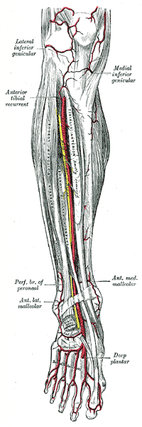
Anterior tibial artery, dorsalis pedis artery and the muscles and bones of the leg - anterior view. (Lat. tarsal very faintly labeled at bottom center.)
Details
Source Dorsalis pedis artery
Identifiers
Latin arteria tarsalis lateralis
TA98 A12.2.16.049
TA2 4715
FMA 44595
Anatomical terminology

The lateral tarsal artery (tarsal artery) arises from the dorsalis pedis, as that vessel crosses the navicular bone; it passes in an arched direction lateralward, lying upon the tarsal bones, and covered by extensor hallucis brevis and extensor digitorum brevis; it supplies these muscles and the articulations of the tarsus, and receives the arcuate over the base of the fifth metatarsal. It may receive contributions from branches of the anterior lateral malleolar and the perforating branch of the peroneal artery directed towards the joint capsule, and from the lateral plantar arteries through perforating arteries of the foot.

Related Research Articles

<span class="mw-page-title-main">Human leg</span> Lower extremity or limb of the human body (foot, lower leg, thigh and hip)

The leg is the entire lower limb of the human body, including the foot, thigh or sometimes even the hip or buttock region. The major bones of the leg are the femur, tibia, and adjacent fibula. The thigh is between the hip and knee, while the calf (rear) and shin (front) are between the knee and foot.

<span class="mw-page-title-main">Radial nerve</span> Nerve in the human body that supplies the posterior portion of the upper limb

The radial nerve is a nerve in the human body that supplies the posterior portion of the upper limb. It innervates the medial and lateral heads of the triceps brachii muscle of the arm, as well as all 12 muscles in the posterior osteofascial compartment of the forearm and the associated joints and overlying skin.

<span class="mw-page-title-main">Anatomical snuffbox</span> Indent on back of hand between tendons

The anatomical snuff box or snuffbox or foveola radialis is a triangular deepening on the radial, dorsal aspect of the hand—at the level of the carpal bones, specifically, the scaphoid and trapezium bones forming the floor. The name originates from the use of this surface for placing and then sniffing powdered tobacco, or "snuff." It is sometimes referred to by its French name tabatière.

<span class="mw-page-title-main">Extensor carpi radialis longus muscle</span>

The extensor carpi radialis longus is one of the five main muscles that control movements at the wrist. This muscle is quite long, starting on the lateral side of the humerus, and attaching to the base of the second metacarpal bone.

In human anatomy, extensor carpi radialis brevis is a muscle in the forearm that acts to extend and abduct the wrist. It is shorter and thicker than its namesake extensor carpi radialis longus which can be found above the proximal end of the extensor carpi radialis brevis.

<span class="mw-page-title-main">Deep femoral artery</span> Large branch of the femoral artery that supplies the thigh

The deep femoral artery also known as the deep artery of the thigh, or profunda femoris artery, is a large branch of the femoral artery. It travels more deeply ("profoundly") than the rest of the femoral artery. It gives rise to the lateral circumflex femoral artery and medial circumflex femoral artery, and the perforating arteries, terminating within the thigh.

<span class="mw-page-title-main">Fibula</span> Leg bone in vertebrates

The fibula or calf bone is a leg bone on the lateral side of the tibia, to which it is connected above and below. It is the smaller of the two bones and, in proportion to its length, the most slender of all the long bones. Its upper extremity is small, placed toward the back of the head of the tibia, below the knee joint and excluded from the formation of this joint. Its lower extremity inclines a little forward, so as to be on a plane anterior to that of the upper end; it projects below the tibia and forms the lateral part of the ankle joint.

In human anatomy, the extensor pollicis longus muscle (EPL) is a skeletal muscle located dorsally on the forearm. It is much larger than the extensor pollicis brevis, the origin of which it partly covers and acts to stretch the thumb together with this muscle.

<span class="mw-page-title-main">Dorsal interossei of the foot</span> Four muscles situated between the metatarsal bones

In human anatomy, the dorsal interossei of the foot are four muscles situated between the metatarsal bones.

<span class="mw-page-title-main">Extensor pollicis brevis muscle</span>

In human anatomy, the extensor pollicis brevis is a skeletal muscle on the dorsal side of the forearm. It lies on the medial side of, and is closely connected with, the abductor pollicis longus. The extensor pollicis brevis (EPB) belongs to the deep group of the posterior fascial compartment of the forearm. It is a part of the lateral border of the anatomical snuffbox.

<span class="mw-page-title-main">Adductor magnus muscle</span> Muscle in the thigh

The adductor magnus is a large triangular muscle, situated on the medial side of the thigh.

<span class="mw-page-title-main">Extensor digitorum brevis muscle</span> Muscle on the upper surface of the foot that helps extend digits 2 through 4

The extensor digitorum brevis muscle is a muscle on the upper surface of the foot that helps extend digits 2 through 4.

<span class="mw-page-title-main">Deep fibular nerve</span> Type of nerve

The deep fibular nerve begins at the bifurcation of the common fibular nerve between the fibula and upper part of the fibularis longus, passes infero-medially, deep to the extensor digitorum longus, to the anterior surface of the interosseous membrane, and comes into relation with the anterior tibial artery above the middle of the leg; it then descends with the artery to the front of the ankle-joint, where it divides into a lateral and a medial terminal branch.

<span class="mw-page-title-main">Perforating arteries</span>

The perforating arteries are branches of the deep artery of the thigh, usually three in number, so named because they perforate the tendon of the adductor magnus to reach the back of the thigh. They pass backward near the linea aspera of the femur underneath the small tendinous arches of the adductor magnus muscle.

The anterior lateral malleolar artery is an artery in the ankle.

The arcuate artery of the foot arises from dorsalis pedis slightly anterior to the lateral tarsal artery, specifically over the naviculocuneiform joint; it passes lateralward, over the bases of the lateral four metatarsal bones, beneath the tendons of the extensor digitorum brevis, its direction being influenced by its point of origin; and it terminates in the lateral tarsal artery. It communicates with the plantar arteries through the perforating arteries of the foot.

<span class="mw-page-title-main">Fascial compartments of arm</span> Anatomical compartments

The fascial compartments of arm refers to the specific anatomical term of the compartments within the upper segment of the upper limb of the body. The upper limb is divided into two segments, the arm and the forearm. Each of these segments is further divided into two compartments which are formed by deep fascia – tough connective tissue septa (walls). Each compartment encloses specific muscles and nerves.

<span class="mw-page-title-main">Radial collateral ligament of wrist joint</span>

The radial collateral ligament extends from the tip of the styloid process of the radius and attaches to the radial side of the scaphoid, immediately adjacent to its proximal articular surface and some fibres extend to the lateral side of the trapezium.

<span class="mw-page-title-main">Outline of human anatomy</span> Overview of and topical guide to human anatomy

The following outline is provided as an overview of and topical guide to human anatomy:

<span class="mw-page-title-main">Extrinsic extensor muscles of the hand</span>

The extrinsic extensor muscles of the hand are located in the back of the forearm and have long tendons connecting them to bones in the hand, where they exert their action. Extrinsic denotes their location outside the hand. Extensor denotes their action which is to extend, or open flat, joints in the hand. They include the extensor carpi radialis longus (ECRL), extensor carpi radialis brevis (ECRB), extensor digitorum (ED), extensor digiti minimi (EDM), extensor carpi ulnaris (ECU), abductor pollicis longus (APL), extensor pollicis brevis (EPB), extensor pollicis longus (EPL), and extensor indicis (EI).

References

PD-icon.svgThis article incorporates text in the public domain from page 637 of the 20th edition of Gray's Anatomy (1918)